Figures & data
Table 1. Primers used in RT-PCR.
Figure 1. SEM photographs from Bio-Oss®-coated polycaprolactone nanofibrous scaffolds fabricated using electrospinning at two magnifications (1.00 kx (a), 5.00 kx (b)).
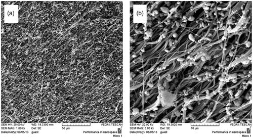
Figure 2. Stem cell images at two magnifications ×10 and ×40, BFP-MSCs (a, b), BM-MSCs (c, d) and USSCs (e, f) at passages two while cultured on tissue culture polystyrene (TCPS) under DMEM supplemented with FBS 10%.
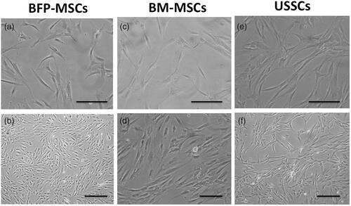
Figure 3. SEM photographs from Bio-Oss®-coated Polycaprolactone nanofibrous scaffolds, two weeks after BFP-MSCs (a, b), BM-MSCs (c, d) and USSC (e, f) seeding under osteogenic differentiation medium at two magnifications.
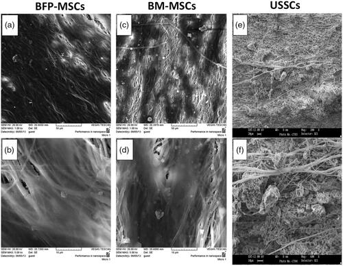
Figure 4. Survival rate of BFP-MSCs, BM-MSCs and USSC while cultured on tissue culture polystyrene (TCPS), polycaprolactone nanofibrous scaffold (PCL) and Bio-Oss®-coated PCL nanofibrous scaffold (PCL-Bio) during days 1, 3, 5 and 7 cultured under DMEM supplemented FBS 10% (asterisks: significant difference between the groups at p < .05).
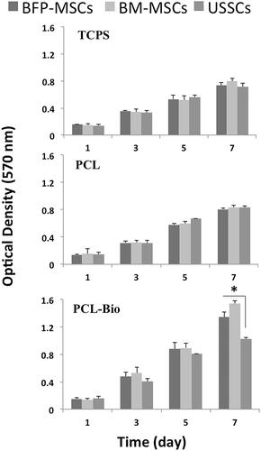
Figure 5. Alizarin red staining of BFP-MSCs (a), BM-MSCs (b) and USSCs (c) two weeks after cultured on tissue culture polystyrene (TCPS) under osteogenic differentiation medium, magnification ×40. Calcium content of the stem cells while cultured on tissue culture polystyrene (TCPS), Polycaprolactone nanofibrous scaffold (PCL) and Bio-Oss®-coated PCL nanofibrous scaffold (PCL-Bio) under osteogenic differentiation medium on day 7 (d) and day 14 (e) (asterisks: significant difference between the groups at p < .05).
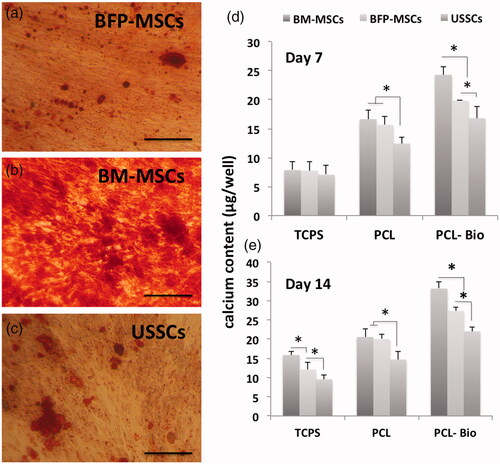
Figure 6. Alkaline phosphatase (ALP) activity of the stem cells while cultured on tissue culture polystyrene (TCPS), polycaprolactone nanofibrous scaffold (PCL) and Bio-Oss®-coated PCL nanofibrous scaffold (PCL-Bio) under osteogenic differentiation medium on day 7 and day 14 (asterisks: significant difference between the groups at p < .05).
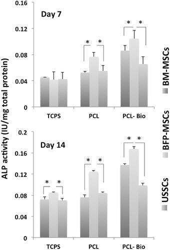
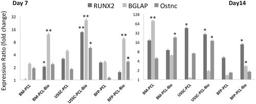
Figure 7. Quantitative expression changes of the Runx2, osteocalcin (BGLAP) and osteonectin in BFP-MSCs, BM-MSCs and USSCs while cultured on polycaprolactone nanofibrous scaffold (PCL) and Bio-Oss®-coated PCL nanofibrous scaffold (PCL-Bio) under osteogenic differentiation medium on days 7 and 14 (asterisks: significant difference between the groups at p < .05).