Figures & data
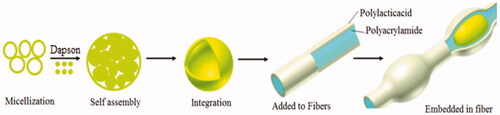
Figure 1. 3D molecular structure of dapson (a) and the XRD patterns of the polyacrylamide/polylactic acid co-assembled core/shell nanofiber structures (b).
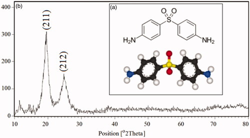
Table 1. Review of different core/shell nanofibers for drug delivery.
Table 2. Reaction conditions for preparation of micro and nanoemulsion/dapson.
Scheme 1. The summary of the experimental method to formation of polyacrylamide/polylactic acid co-assembled core/shell nanofibers contains dapson nanoemulsion.
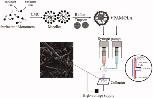
Figure 2. SEM image cumulative of dapsone microemulsion (a), polyacrylamide/polylactic acid core/shell nanofibers (b) dapsone nanoemulsion loaded in polyacrylamide/polylactic acid as core/shell nanofibers (c).
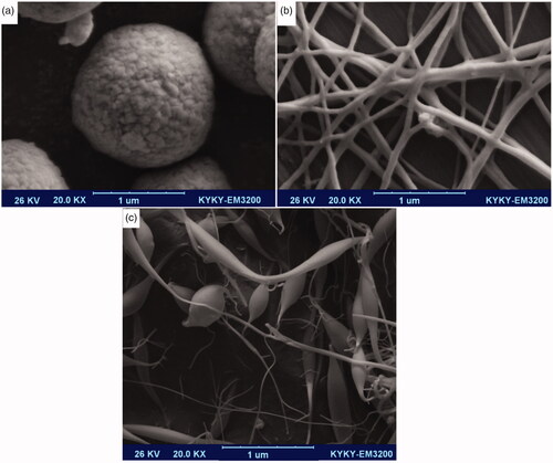
Figure 3. DLS analysis of dapsone microemulsion (a), polyacrylamide/polylactic acid core/shell nanofibers (b) dapsone nanoemulsion loaded in polyacrylamide/polylactic acid as core/shell nanofibers (c).
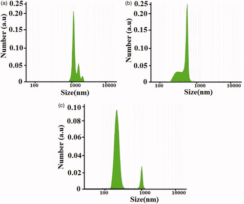
Figure 4. TEM images of the synthesized dapsone micro and nanoemulsion (a) and drug loaded on polyacrylamide/polylactic core/shell nanofibers (b).
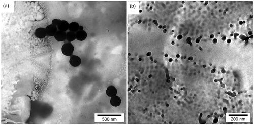
Figure 5. TGA and DTG curves (a) and AFM images of the synthesized for dapsone micro and nanoemulsion drug loaded in polyacrylamide/polylactic core/shell nanofibers (b).
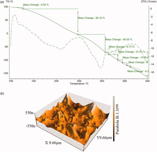
Figure 6. The relationship between advancing contact angle (θA) and duration of different drop volumes on surface (a) and N2 adsorption–desorption isotherms of polyacrylamide/polylactic acid core/shell nanofibers (b).
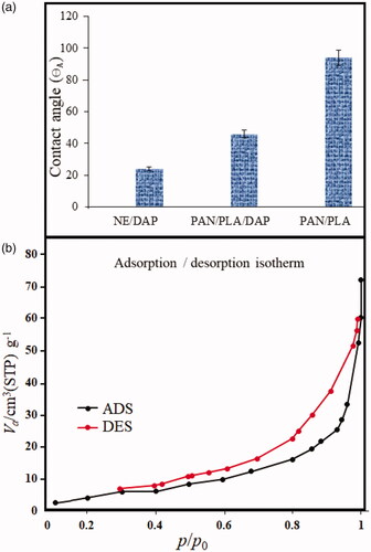
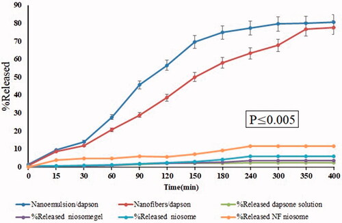
Figure 7. The cumulative release profiles of dapsone from nanoemulsion and core/shell nanofibers modified with nanoemulsion. Percentage of released niosomegel, dapsone solution, noisome and nanofiber niosomes.