Figures & data
Figure 1. UV–visible spectrum absorption pattern of gold nanoparticles synthesised from Strychni semen.
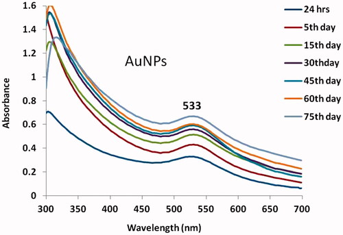
Figure 2. HR-Transmission electron microscopy (TEM) & Energy dispersive X-ray analysis (EDX) of gold nanoparticles synthesised from Strychni semen.
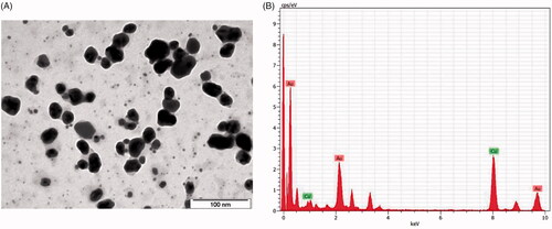
Figure 3. Fourier-transform infrared spectroscopy analysis and SAED pattern of gold nanoparticles synthesised from Strychni semen.
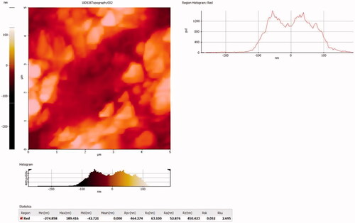
Figure 4. Atomic force microscopyanalysis of gold nanoparticles synthesised from Strychni semen.
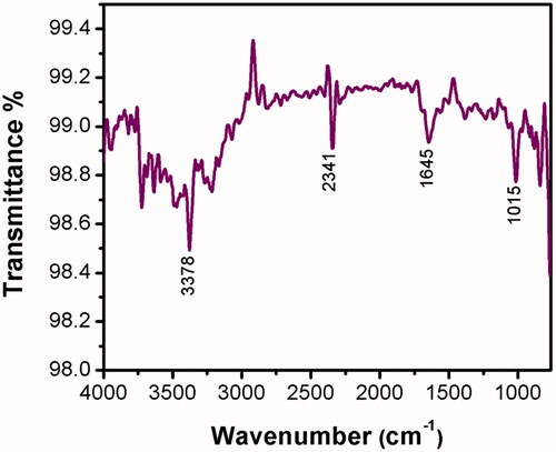
Figure 5. Cytotoxicity effect of gold nanoparticles synthesised from Strychni semen in cholangiocarcinoma cell (KMCH-1). The number of viable cells after treatment is expressed as a percentage of the vehicle-only control. This experiment was repeated thrice and the bars in the graph represent S.E. (*p < .05).
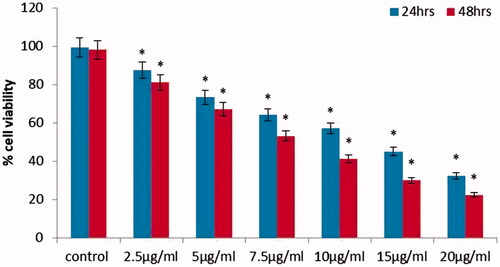
Figure 6. Apoptotic effect of gold nanoparticles synthesised from Strychni semen in KMCH-1 Cells.
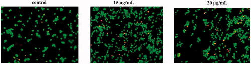
Figure 7. Intracellular production of ROS levels in KMCH-1 cells. This experiment was repeated thrice and the bars in the graph represent S.E. (*p < .05).
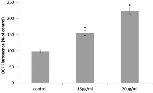
Figure 8. Caspase-3, Caspase-9 activity in KMCH-1 cells. This experiment was repeated thrice and the bars in the graph represent S.E. (*p < .05, #p < .01).
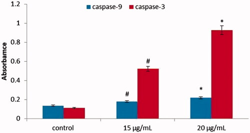
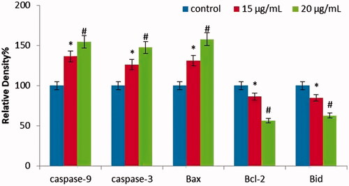
Figure 9. Anticancer effect of gold nanoparticles synthesised from Strychni semen on apoptotic gene expression in KMCH-1 Cells. This experiment was repeated thrice and the bars in the graph represent S.E. (*p < .05, #p < .01).