Figures & data
Figure 1. Histamine H1 receptor (H1R) is expressed in pre-osteoblast MC3T3-E1 cells. (A) RT-PCR results revealed that H1R is expressed in MC3T3-E1 cells at the gene level; (B) Western blot analysis revealed that H1R is expressed in MC3T3-E1 cells at the protein level. B16-F10 cells were used as an important positive control.
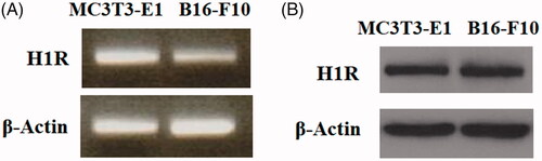
Figure 2. Expression of Histamine H1 receptor (H1R) was increased during osteoblast differentiation process of MC3T3-E1 cells. Pre-osteoblast MC3T3-E1 cells were cultured with osteogenic medium (OM) for various time durations (0, 3, 7 and 14 days). (A) Real-time PCR analysis of H1R; (B) Western blot analysis of H1R (*, #, $, P < .01 vs. previous column group).
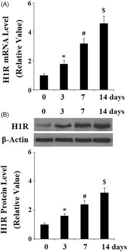
Figure 3. Antagonism of Histamine H1 receptor (H1R) using Loratadine prevented the differentiation and mineralization of MC3T3-E1 cells. MC3T3-E1 cells were cultured with osteogenic medium (OM) in the presence or absence of Loratadine at 50 µM for 14 days. (A) Loratadine reduced OM-induced increase in ALP activity; (B) Alizarin Red staining assay revealed that Loratadine prevented OM-induced mineralization of MC3T3-E1 cells; (C) Real-time PCR analysis demonstrated that Loratadine reduced the gene expression of ALP, OCN, Osx, and Col-I (*, #, P < .01 vs. previous column group).
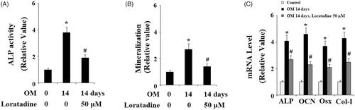
Figure 4. Antagonism of Histamine H1 receptor (H1R) using Loratadine reduced the expression of RUNX-2 during osteoblast differentiation process of MC3T3-E1 cells. Pre-osteoblast MC3T3-E1 cells were treated with osteogenic medium (OM) in the presence or absence of Loratadine (50 µM). (A) Real-time PCR analysis of RUNX-2; (B) Western blot analysis of RUNX-2 (*, #, P < .01 vs. previous column group).

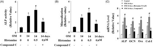
Figure 5. Histamine promoted the differentiation and mineralization of MC3T3-E1 cells. MC3T3-E1 cells were cultured with osteogenic medium (OM) in the presence or absence of Histamine at 0.5 µM for 14 days. (A). Histamine promoted OM-induced increase in ALP activity; (B). Alizarin Red staining assay revealed that Histamine promoted OM-induced mineralization of MC3T3-E1 cells; (C). Real-time PCR analysis demonstrated that Histamine promoted the gene expression of ALP, OCN, Osx, and Col-I (*, #, P < .01 vs. previous column group).
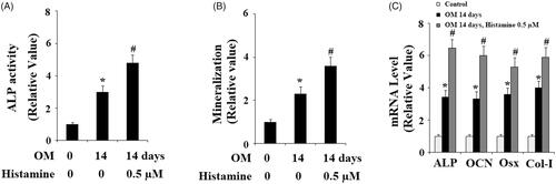
Figure 6. Histamine promoted the expression of RUNX-2 during osteoblast differentiation process of MC3T3-E1 cells. Pre-osteoblast MC3T3-E1 cells were treated with osteogenic medium (OM) in the presence or absence of Histamine at 0.5 µM. (A). Real time PCR analysis of RUNX-2; (B). Western blot analysis of RUNX-2 (*, #, P < .01 vs. previous column group).
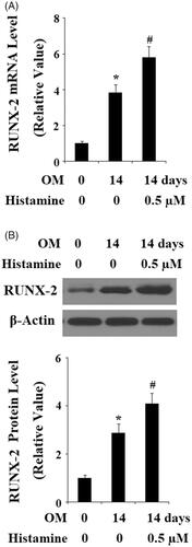
Figure 7. Activation of H1R by Histamine promoted the phosphorylation of eNOS and the production of nitric oxide (NO). (A) Pre-osteoblast MC3T3-E1 cells were treated with Histamine (0.5 µM) for 15 min, 30 min, and 1 h, phosphorylated and total levels of eNOS were determined by western blot analysis; (B) Pre-osteoblast MC3T3-E1 cells were treated with Histamine (0.5 µM) for 1 h, 3 h, and 6 h, production of NO was determined by DAF FM DA; (C) Pre-osteoblast MC3T3-E1 cells were treated with Histamine (0.5 μM) for 15 min, 30 min, and 1 h, phosphorylated and total levels of AMPK were determined by western blot analysis (*, #, $, P < .01 vs. previous column group).
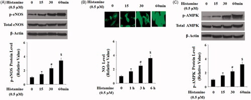
Figure 8. The effects of H1R activation by Histamine on the differentiation and mineralization of MC3T3-E1 cells were suppressed by the specific AMPK inhibitor compound C. MC3T3-E1 cells were maintained in osteogenic medium in the absence or presence of Histamine (0.5 μM) or compound C for 14 days. (A) ALP activity; (B) Quantification of Alizarin Red S staining; (C) Gene expression of ALP, OCN, Osx, and Col-I (*, #, $, P < .01 vs. previous column group).