Figures & data
Figure 1. DPP-4 is expressed in human dental pulp cells. Human umbilical vein endothelial cells (HUVECs) were used as a positive control. (A). Reverse transcription PCR (RT-PCR) analysis of DPP-4; (B). Western blot analysis of DPP-4.
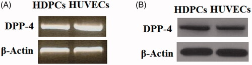
Figure 2. Lipopolysaccharide (LPS) treatment increased the expression of DPP-4 in human dental pulp cells. Human dental pulp cells were treated with LPS at the concentrations of 50, 100, 150 ng/ml for 24 h. (A). mRNA levels of DPP-4 were determined by real time PCR analysis; (B). Protein levels of DPP-4 were determined by western blot analysis (*, #, $ p < .01 vs. previous column group).
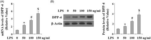
Figure 3. Saxagliptin ameliorated LPS-induced oxidative stress in human dental pulp cells. Human dental pulp cells were treated with 100 ng/ml LPS in the presence or absence of saxagliptin (500 nM, 1 μM) for 24 h. (A). Production of reactive oxygen species (ROS); (B). Intracellular levels of GSH (*, #, $ p < .01 vs. previous column group).
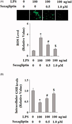
Figure 4. Saxagliptin ameliorated LPS-induced mitochondrial dysfunction in human dental pulp cells. Human dental pulp cells were treated with 100 ng/ml LPS in the presence or absence of saxagliptin (500 nM, 1 μM) for 48 h. (A). MMP was determined by TMRM; (B). Intracellular ATP (*, #, $ p < .01 vs. previous column group).
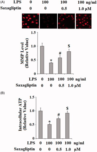
Figure 5. Saxagliptin prevented LPS-induced cell death of human dental pulp cells. Human dental pulp cells were treated with 100 ng/ml LPS in the presence or absence of saxagliptin (500 nM, 1 μM) for 48 h. (A). Cell viability was determined by MTT assay; (B). LDH release (*, #, $ p < .01 vs. previous column group).
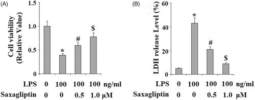
Figure 6. Saxagliptin ameliorated LPS-induced pro-inflammatory cytokines in human dental pulp cells. Human dental pulp cells were treated with 100 ng/ml LPS in the presence or absence of saxagliptin (500 nM, 1 μM) for 48 h. (A). TNF-α, IL-1β, IL-8 at the mRNA levels were determined by real time PCR analysis; (B). TNF-α, IL-1β, IL-8 at the protein levels were determined by western blot analysis (*, #, $ p < .01 vs. previous column group).
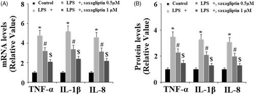
Figure 7. Saxagliptin inhibited LPS-induced activation of p38 in human dental pulp cells. Human dental pulp cells were treated with 100 ng/ml LPS in the presence or absence of saxagliptin (500 nM, 1 μM) for 2 h. Phosphorylated and total levels of p38 were determined by western blot analysis (*, #, $ p < .01 vs. previous column group).
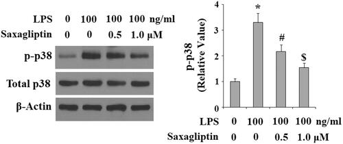
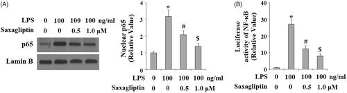
Figure 8. Saxagliptin inhibited LPS-induced activation of NF-κB. Human dental pulp cells were treated with 100 ng/ml LPS in the presence or absence of saxagliptin (500 nM, 1 μM) for 24 h. (A). Nuclear translocation of p65; (B). Luciferase activity of NF-κB (*, #, $ p < .01 vs. previous column group).