Figures & data
Figure 1. miR-384 was down-regulated in (A) clinical human osteosarcoma tissues and (B) osteosarcoma cell lines. miR-384 was quantified using qRT-PCR. Means ± SD were shown. **p < .01, ***p < .001.
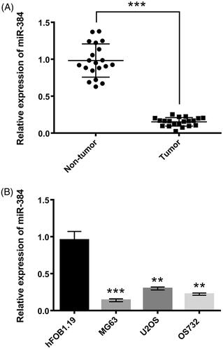
Figure 2. miR-384 inhibited growth and metastasis of MG63 cells. (A) Relative miR-384 expression was determined after transfection assay. miR-384 was examined using qRT-PCR. miR-384 (B) inhibited cell viability, (C) migration and (D) invasion, as well as (E) promoted apoptosis and (F) influenced apoptosis-related proteins. However, knocking down miR-384 showed the contrary effects. Cell viability was assessed by CCK-8; Migration and invasion were evaluated by transwell assay; Apoptotic cells were observed with flow cytometer; Proteins involved in apoptosis progression was detected using Western blotting. Means ± SD were shown. *p < .05, **p < .01.
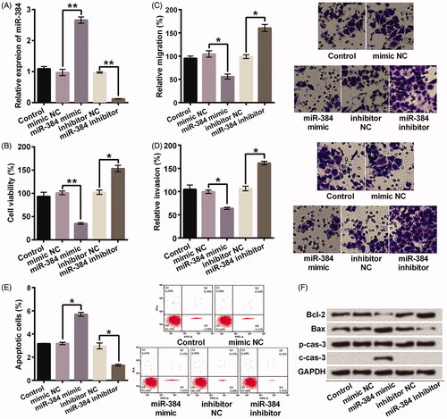
Figure 3. SLBP was a direct target of miR-384. SLBP expression was negatively regulated by miR-384 at (A) mRNA and (B) protein levels. mRNA and protein were examined using qRT-PCR and Western blotting, respectively. (C) Luciferase activities of reporter constructs containing wild-type or mutant 3′-UTR of SLBP were assayed and normalized to those of Renilla. Means ± SD were shown. *p < .05, **p < .01.
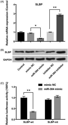
Figure 4. miR-384 silence promoted the growth and metastasis of MG63 cells by elevating expression of SLBP. Transfection of si-SLBP inhibited SLBP expression in miR-384 silenced cells at (A) mRNA (B) and protein levels. SLBP was determined at both mRNA and protein levels using qRT-PCR and Western blotting. SLBP silence abolished the promoting effects of miR-384 silence on (C) cell viability, (D) cell migration and (E) invasion. SLBP silence reduced (F) the inhibitory effect of miR-384 silence on apoptosis and (G) the effect on apoptosis-related proteins. CCK-8 assay was applied for cell viability; Transwell assay was conducted to examine migration and invasion behaviours; Apoptotic cells were observed under a flow cytometer; Western blotting assay was used to quantify protein associated with apoptosis. Means ± SD were shown. *p < .05, **p < .01.
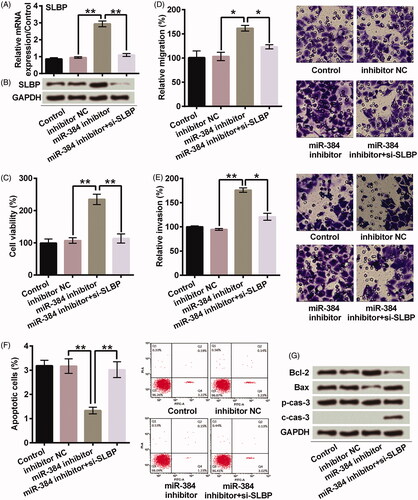
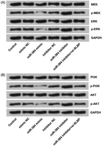
Figure 5. miR-384 silence activated (A) MEK/ERK and (B) PI3K/AKT signalling pathways in MG63 cells by up-regulating SLBP. However, miR-384 overexpression inhibited the activations of both pathways. MEK, ERK, PI3K and AKT, as well as their phosphorylated forms, were analyzed using Western blotting assay.