Figures & data
Figure 1. Primary BMSCs (A) at 3 days and passage BMSCs (B) at 7 days.
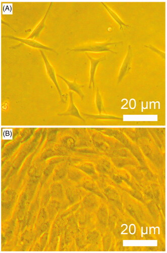
Table 1. Comparisons of chondrocyte amounts, absorbance of MTT assay, and glycosaminoglycan contents between chondrocyte induction group and control group.
Figure 2. Schematic (A) and experimental setup (B) of a custom-design bioreactor system. It consists of bioreactor vessel, peristaltic pump, air filter, two tubes (tube 1 and tube 2), three way valve and media bottle. These parts are indicated in the figure. It is located in a humidity-saturated incubator with 5% CO2 at 37 °C and is used for cells-loaded scaffold culture.
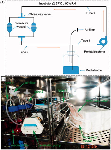
Figure 3. SEM images of cells on bioceramic scaffolds. (A) Chondrocytes on β-TCP bioceramic scaffolds. (B) Osteoblasts on β-TCP bioceramic scaffolds. (C) Section image of β-TCP bioceramic scaffolds. (D) Surface image of β-TCP bioceramic scaffolds.
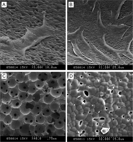
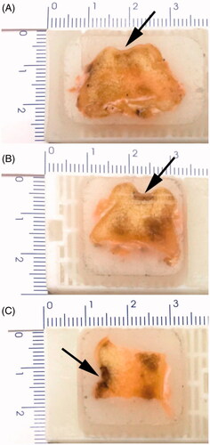
Figure 4. General observation of articular cartilage defects (A), implantation of bioceramic scaffolds (B), and articular cartilage regeneration after 12 weeks postimplantation (C) in Beagle femoral trochleae. (a): chondrocyte/osteoblast-loaded β-TCP bioceramic scaffold group, (b): chondrocyte-loaded β-TCP bioceramic scaffold group, and (c): β-TCP bioceramic scaffold group. n = 3.
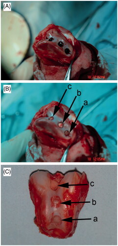
Figure 5. Histological observation of Beagle femoral trochleae after 12 weeks postimplantation. (A) Chondrocyte/osteoblast-loaded β-TCP bioceramic scaffold group. (B) Chondrocyte-loaded β-TCP bioceramic scaffold group. (C) β-TCP bioceramic scaffold. Arrows indicate the cartilage defects where the scaffolds were implanted.