Figures & data
Figure 1. Expression of miR-10b in both CRC tissues and CRC cell lines. (A) miR-10b was overexpressed in metastatic CRC samples (n = 14), compared with non-metastatic ones (n = 16). (B) The expression of miR-10b in 6 CRC cell lines and normal mucosa cells was different from each other. Cell results shown are representative of three independent experiments. Data are given as means ± SD.
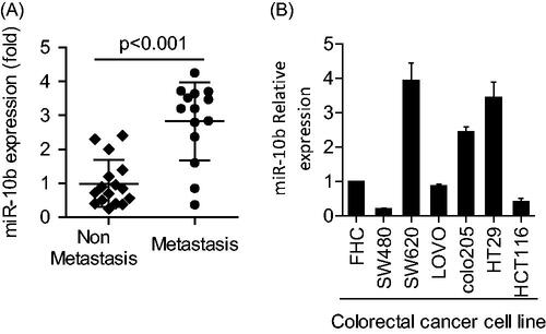
Figure 2. Effects of miR-10b on metastasis, proliferation, apoptosis and cell-cycle of SW620 cells. (A) the expression of E-cadherin increased after treatment of miR-10b inhibitor, but the expression of cyclin D1 and Snail protein decreased. (B,C) Downregulation of miR-10b notably inhibited cell migration and invasion of SW620 cells (100× magnification). (D) Transfecting miR-10b inhibitors significantly reduced proliferation of SW620 cells (*p < .05). (E) miR-10b inhibition leads to cell-cycle arrest in SW620 cells. Cell cycle was stained with PI and analyzed by flow cytometer after 2 days following transfections in SW620 cells. (F) Inhibition of miR-10b expression enhanced apoptosis. Cells were stained with PI and Annexin V staining, and analyzed by flow cytometer after 2 days following transfections. Cell results shown are representative of three independent experiments. Data are given as means ± SD.
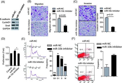
Figure 3. The KLF4 3’-UTR is a target of miR-10b. (A,B) KLF4-encoded mRNA contains a 3`- UTR which perfectly matches to the seed region of miR-10b. Plasmid with the full-length KLF4-3’- UTR insert and plasmid with a mutant KLF4-3`- UTR (KLF4-3`UTR-mut). (B) klf4 mRNA was detected by qPCR after the treatment of miR-10b inhibitor in SW620. (C) KLF4 expression was detected by western blot after the treatment of miR-10b inhibitor in SW620 and HT29. (D) The luciferase activity was measured with transfection of miR-10b inhibitor and KLF4-3`UTR or KLF4-3`UTR-mut plasmid in HT29. (E) The luciferase activity was measured with transfection of miR-10b mimic and KLF4-3`UTR or KLF4-3`UTR-mut plasmid in HEK293T cell line. Renilla luciferase activity was normalized to Firefly luciferase activity. Cell results shown are representative of three independent experiments. Data are given as means ± SD.
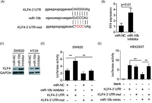
Figure 4. Effects of KLF4 on metastasis, proliferation, apoptosis and cell cycle of SW620 cell lines. (A) Over-expression of KLF4 greatly inhibited cell migration and invasion of SW620 cells (100× magnification). (B) Transfecting KLF4 plasmids significantly reduced proliferation of SW620 cells (*P < .05). (C) Over-expression of KLF4 leads to cell-cycle arrest in SW620 cells. (D) Over-expression of KLF4 enhanced apoptosis. SW620 cells were analyzed with PI and Annexin V staining by flow cytometer after 3 days following transfections. Cell results shown are representative of three independent experiments. Data are given as means ± SD.
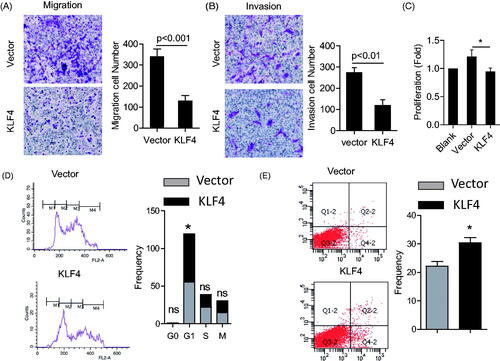
Figure 5. KLF4 Partly attenuated the function of miR-10b. SW620 cells were transfected with siRNA for KLF4 mRNA and miR-10b inhibitor. (A) KLF4, E-cadherin, cyclinD1 and Snail expression were detected by western blot. (B–D) Then migration and invasion assays were evaluated by Transwell and the proliferation was detected by MTT. Cell results shown are representative of three independent experiments. Data are given as means ± SD.
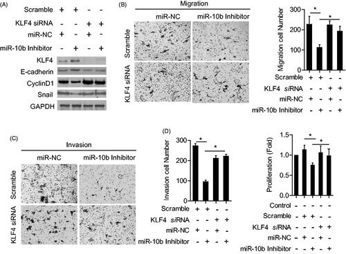
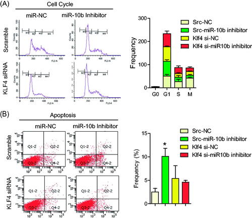
Figure 6. KLF4 Partly attenuated the function of miR-10b on cell cycle and apoptosis of CRC cells. SW620 cells were transfected with siRNA for KLF4 mRNA and miR-10b inhibitor. Then migration and invasion assays were performed to evaluate the effect of KLF4 on the function of miR-10b. (A,B) cell cycle and (C,D) apoptosis assays were also conducted. Cell results shown are representative of three independent experiments. Data are given as means ± SD.