Figures & data
Figure 1. Overview of preparation method of an ECM hydrogel from human adipose tissue. SVF: stromal vascular fraction; ECM: extracellular matrix.
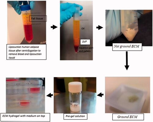
Figure 2. Representative data of CD‐surface marker expression in immortalized ASCs.
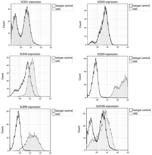
Figure 3. Immortalized ASC retain cell differentiation capacity to adipocytes, osteoblast, smooth muscle cells. (A) Immortalized ASC differentiated to adipocytes. (B) Differentiated to osteoblasts. (C) Differentiated to smooth muscle cells.
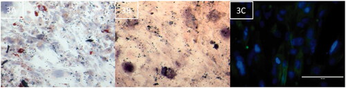
Figure 4. Optical density plotted against the serially diluted CMe from human adipose tissue-derived hydrogels after four different time points (log scale). PK84 fibroblasts were treated with hydrogel CMe or controls for 48 h. Puromycin was used as positive control. Normal culture medium was used as negative control. Results are presented as mean with standard error of the mean of triplicates of three independent donors. CMe: conditioned medium.
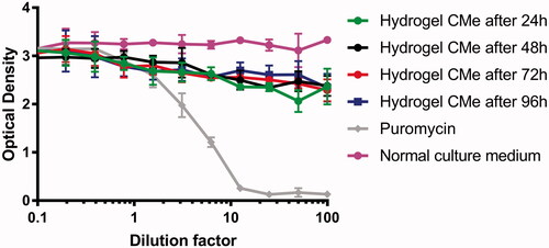
Figure 5. (A,C,E) Representative fluoromicrographs of iASC13EGFP cultured on top of human adipose tissue-derived ECM hydrogels after respectively 1, 6 and 6 days. (B,D,F) Representative fluoromicrographs of iASC13EGFP cultured on tissue culture plastic (control) for respectively 1, 6 and 6 days. Scale bar: A–D: 400 µm, E–F: 200 µm. iASC13EGFP: immortalized EGFP-tagged adipose derived stromal cells; ECM: extracellular matrix.
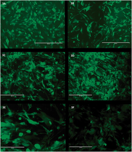
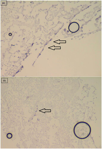
Figure 6. Hematoxylin-stained sections of iASC13EGFP cultured on top of human adipose tissue-derived ECM hydrogels after 6 days. Note that cells (visible by their dark blue nuclei) have migrated into the gel (arrows) at the periphery (A) and to the centre (B). iASC13EGFP: immortalized EGFP-tagged adipose derived stromal cells; ECM: extracellular matrix.