Figures & data
Figure 1. Propofol suppresses cell proliferation and metastasis in JEG-3 cells. JEG-3 cells were treated with various concentration of propofol (10 nM, 100 nM, 1 μM and 10 μM). (A) Cell viability was assessed by CCK-8 assay. (B) Relative migration and (C) invasion was measured using Transwell analysis. (D) The expression of EMT-related factors in JEG-3 cells was detected using western blot. (E) Western blot analysis of cell proliferation-regulated factors in JEG-3 cells. *p < .05, **p < .01, ***p < .001.
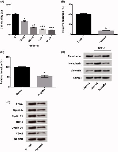
Figure 2. Propofol up-regulates miR-495 expression in JEG-3 cells. After treated with propofol (1 μM), the expression of miR-495 in JEG-3 cells was detected using qRT-PCR. **p < .01.
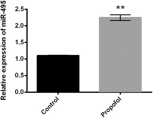
Figure 3. Suppression of miR-495 blocks the regulation of propofol in cell proliferation and metastasis in JEG-3 cells. JEG-3 cells were transfected with miR-495 inhibitor and the corresponding negative control and then were treated with propofol (1 μM). (A) The expression of miR-495 was measured using qRT-PCR. Then, (B) cell viability, (C) cell proliferation, (D) relative migration and (E) invasion, and (F) the expression of EMT-regulated core factors were respectively detected using CCK-8 assay, Ki67+ assay, Transwell analysis, and western blot analysis. *p < .05, **p < .01.
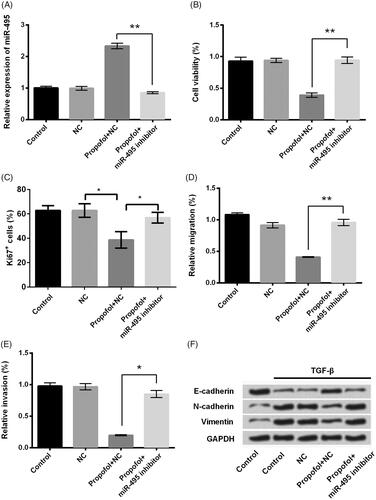
Figure 4. Propofol decreases AKT1 and Twist1 expression through regulating miR-495. JEG-3 cells were transfected with miR-495 inhibitor and the corresponding negative control and then were treated with propofol (1 μM). Then, the mRNA and protein expression of (A) AKT1 and (B) Twist1 was evaluated using qRT-PCR and western blot analysis. *p < .05, **p < .01.
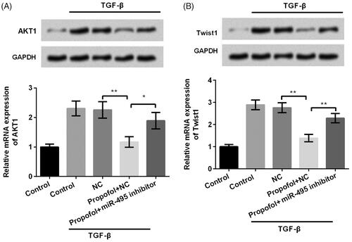
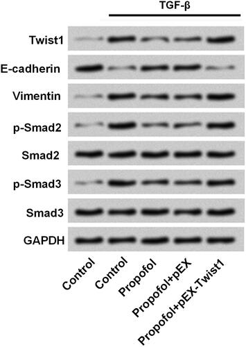
Figure 5. Twist1 participates in the regulation of propofol on TGF-β signalling inactivation in JEG-3 cells. JEG-3 cells were transfected with Twist1 overexpress-vector and the corresponding negative control. The protein expressions of Twist1, E-cadherin, Vimentin, p-Smad2, Smad2, p-Smad3 and Smad3 were measured by western blot.