Figures & data
Figure 1. Secretion of Amyloid β (Aβ) was increased in AD cell models. The levels of secreted Aβ40 and Aβ42 as determined by an ELISA assay (a, p < .01 vs. vehicle control).
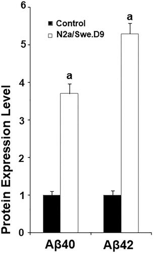
Figure 2. Anagliptin (0.5, 1 μM) ameliorates oxidative stress in N2a/Swe.D9 cells. Cells were incubated with 0.5, 1 μM anagliptin for 24 h. (A) ROS was measured through DCFH-DA staining; (B) Cells were stained with dihydroethidium (DHE) (a, b, c, p < .01 vs. the previous group).
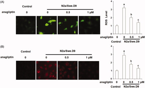
Figure 3. Anagliptin (0.5, 1 μM) suppresses the expression of NOX-4. Cells were incubated with 0.5, 1 μM anagliptin for 24 h. (A) mRNA of NOX-4; (B) Protein of NOX-4 (a, b, c, p < .01 vs. the previous group).
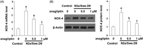
Figure 4. Anagliptin (0.5, 1 μM) restored the reduction of reduced glutathione (GSH) in AD cell models. N2a/Swe.D9 cells were stimulated with 0.5, 1 μM anagliptin for 24 h. (A) Reduced GSH was determined; (B) GPx activity was measured (a, b, c, p < .01 vs. the previous group).
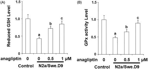
Figure 5. Anagliptin (0.5, 1 μM) improved mitochondrial dysfunction in an AD cell model. N2a/Swe.D9 cells were cultured with 0.5, 1 μM anagliptin for 24 h. (A) Intracellular levels of MMP were measured by TMRM staining; (B) Intracellular ATP was measured using a bioluminescence method (a, b, c, p < .01 vs. the previous group).
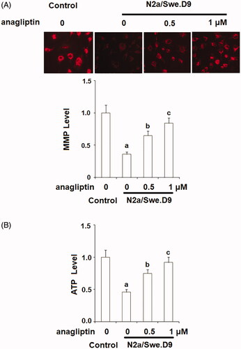
Figure 6. Anagliptin (0.5, 1 μM) protects N2a/Swe.D9 cells against secondary insult by H2O2. Cells were stimulated with H2O2 (100 μM) with or without anagliptin (0.5, 1 μM) for 24 h. (A). Cell viability as determined by MTT assay; (B). Leakage of LDH as examined using a kit (a, b, c, p < .01 vs. the previous group).
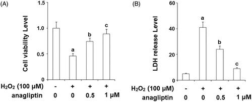
Figure 7. Anagliptin (0.5, 1 μM) protects against H2O2-induced HMGB-1 secretion in an AD cell model. N2a/Swe.D9 cells were stimulated with H2O2 (100 μM) with or without anagliptin (0.5, 1 μM) for 24 h. Secretion of HMGB-1 was measured by ELISA (a, b, c, p < .01 vs. the previous group).
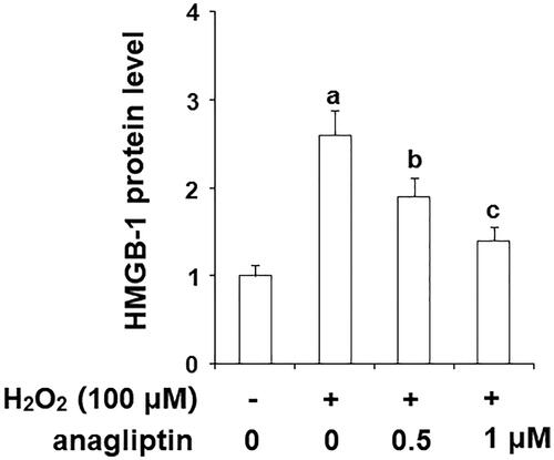
Figure 8. Anagliptin protects against H2O2-induced apoptosis in an AD cell model. N2a/Swe.D9 cells were stimulated with H2O2 (100 μM) with or without anagliptin (0.5, 1 μM) for 24 h. Apoptosis of cells was assayed by TUNEL staining (a, b, c, p < .01 vs. the previous group).
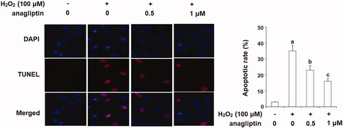
Figure 9. Anagliptin prevents the leakage of cytochrome C from mitochondria into cytosol and activation of caspase-3. N2a/Swe.D9 cells were stimulated with H2O2 (100 μM) with or without anagliptin (0.5, 1 μM) for 24 h. (A) Levels of cytochrome C in the cytosol. (B) Cleaved caspase-3 (a, b, c, p < .01 vs. the previous group).
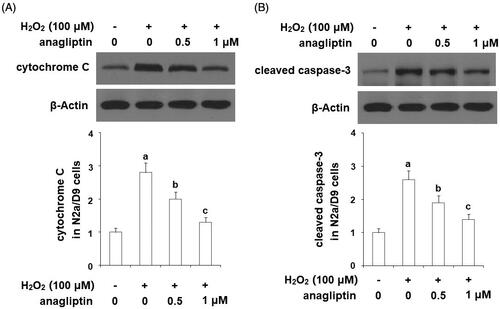
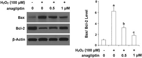
Figure 10. The effects of anagliptin on Bax/Bcl-2 expression. N2a/Swe.D9 cells were stimulated with H2O2 (100 μM) with or without anagliptin (0.5, 1 μM) for 24 h. (A) Representative bands of Bax/Bcl-2; (B) Quantification of Bax/Bcl-2 (a, b, c, p < .01 vs. previous column group).