Figures & data
Figure 1. P2Y11R is expressed on primary human aortic endothelial cells (HAECs). Human huh-7 cells were used as a positive control. (A) RT-PCR of P2Y11R; (B) Western blot analysis of P2Y11R.
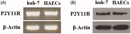
Figure 2. Ox-LDL increases the expression of endothelial P2Y11R in a dose-dependent manner. HAECs were treated with ox-LDL at the concentrations of 0, 5, 10 and 20 mg/L for 24 h. (A) Real-time PCR analysis of P2Y11R; (B) Western blot analysis of P2Y11R (*, #, $, p < .01 vs. vehicle group).
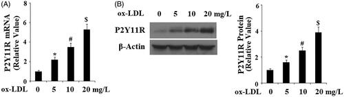
Figure 3. The P2Y11R antagonist NF157 attenuates ox-LDL-induced adhesion of THP-1 monocytes to HAECs. HAECs were treated with 10 mg/L ox-LDL with or without NF157 (25, 50 μM) for 24 h. (A) Representative images of adhesion of THP-1 cells to HAECs; (B) Quantification of adhesive THP-1 cells (*, #, $, p < .01 vs. previous column group).
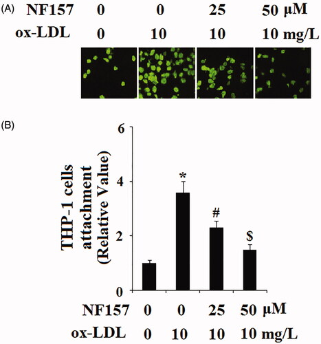
Figure 4. The P2Y11R antagonist NF157 inhibits ox-LDL-induced expression of E-selectin and VCAM-1. HAECs were treated with 10 mg/L ox-LDL with or without NF157 (25, 50 μM) for 24 h. (A) Real-time PCR analysis of E-selectin and VCAM-1; (B). Western blot analysis of E-selectin and VCAM-1 (*, #, $, p < .01 vs. previous column group).
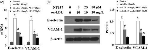
Figure 5. NF157 inhibits ox-LDL-induced oxidative stress. HAECs were treated with 10 mg/L ox-LDL with or without NF157 (25, 50 μM) for 24 h. (A) Production of reactive oxygen species (ROS); (B) Expression of NOX-4 at the mRNA level; (C) Expression of NOX-4 at the protein level (*, #, $, p < .01 vs. previous column group).
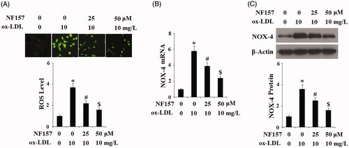
Figure 6. NF157 inhibits ox-LDL-induced expression of the pro-inflammatory cytokines IL-6 and TNF-α. HAECs were treated with 10 mg/L ox-LDL with or without NF157 (25, 50 μM) for 24 h. (A) Real-time PCR analysis of IL-6 and TNF-α at the mRNA level; (B) ELISA analysis of IL-6 and TNF-α at the protein level (*, #, $, p < .01 vs. previous column group).
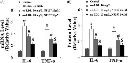
Figure 7. NF157 inhibits ox-LDL-induced activation of MAPK kinase p38. HAECs were treated with 10 mg/L ox-LDL with or without NF157 (25, 50 μM) for 2 h. Phosphorylated and total p38 were determined using western blot analysis (*, #, $, p < .01 vs. previous column group).
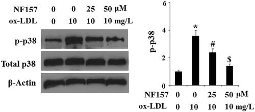
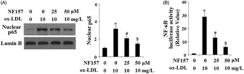
Figure 8. NF157 inhibits ox-LDL-induced activation of NF-κB. HAECs were treated with 10 mg/L ox-LDL with or without NF157 (25, 50 μM) for 24 h. (A) Nuclear level of NF-κB protein p65. Lamin B1 was used as the loading control of nuclei; (B) NF-κB promoter luciferase activity change (*, #, $, p < .01 vs. previous column group).