Figures & data
Figure 1. The effects of paeonol on cell viability in the chondrogenic cell line ATDC5 cells. (A) Molecular structure of paeonol, 2′-hydroxy-4′-methoxyacetophenone. (B) ATDC5 cells were stimulated with 0, 0.1, 1, 10, 50, 100 and 500 μM paeonol for 48 h. Viability of ATDC5 cells was measured by MTT assay. Note: Since 500 μM paeonol reduced cell viability, doses of 50 and 100 μM paeonol were used in the following experiments (*, P < .01 vs. vehicle control).
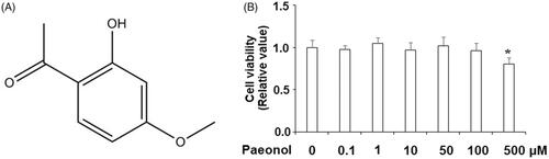
Figure 2. Paeonol reduces inflammatory mediators in chondrogenic cell line ATDC5 cells. ATDC5 cells were incubated with 10 ng/mL IL-1β with or without paeonol (50, 100 μM) for 24 h. (A) mRNA of NOX2 synthase; (B) mRNA of PTGS2; (C) mRNA of NUCB2/nesfatin-1 (*, #, $, P < .01 vs. previous control group).
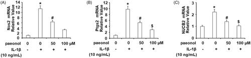
Figure 3. Paeonol inhibits the expression and secretion of pro-inflammatory cytokines in chondrogenic cell line ATDC5 cells. ATDC5 cells were incubated with 10 ng/mL IL-1β with or without paeonol (50, 100 μM) for 24 h. (A) mRNA levels of TNF-α and IL-6; (B) Secretions of TNF-α and IL-6 (*, #, $, P < .01 vs. previous control group).
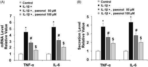
Figure 4. Paeonol inhibits the expression of adhesion molecules. ATDC5 cells were incubated with 10 ng/mL IL-1β with or without paeonol (50, 100 μM) for 24 h. (A) mRNA levels of ICAM-1 and VCAM-1; (B) Protein levels of ICAM-1 and VCAM-1 (*, #, $, P < .01 vs. previous control group).
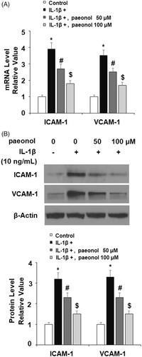
Figure 5. Paeonol suppresses the expression of matrix metalloproteinases in chondrogenic cell line ATDC5 cells. ATDC5 cells were incubated with 10 ng/mL IL-1β with or without paeonol (50, 100 μM) for 24 h. (A) mRNA levels of MMP-3 and MMP-13; (B) Elisa results of MMP-3 and MMP-13 (*, #, $, P < .01 vs. previous control group).
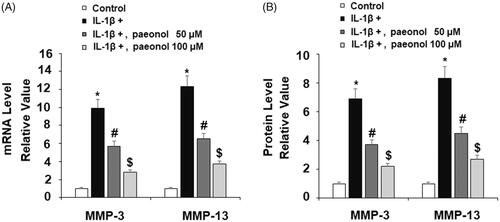
Figure 6. Paeonol ameliorates reduction of type II collagen in chondrogenic cell line ATDC5 cells. ATDC5 cells were incubated with 10 ng/mL IL-1β with or without paeonol (50, 100 μM) for 24 h. Protein level of type II collagen was measured (*, #, $, P < .01 vs. previous control group).
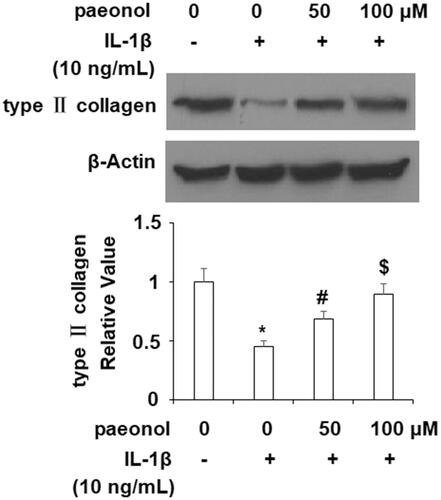
Figure 7. Paeonol inhibits the phosphorylation and degradation of IκBα. ATDC5 cells were incubated with 10 ng/mL IL-1β with or without paeonol (50, 100 μM) for 24 h. Phosphorylated and total IκBα was measured (*, #, $, P < .01 vs. previous control group).
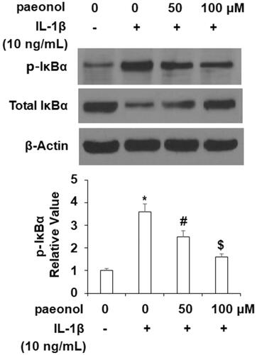
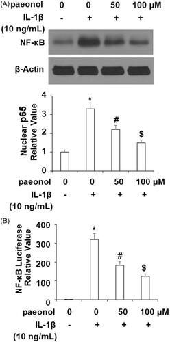
Figure 8. Paeonol suppresses activation of NF-κB. ATDC5 cells were incubated with 10 ng/mL IL-1β with or without paeonol (50, 100 μM) for 24 h. (A) Nuclear level of p65; (B) Luciferase activity of NF-κB (*, #, $, P < .01 vs. previous control group).