
Figures & data
Table 1. Correlation of miRNA-34b expression with the clincopathological characteristics of the cervical cancer patients.
Figure 1. MiR-34b was lower in advanced cervical cancer tissues. (A) MiR-34b was lower in high FIGO status. (B) MiR-34b was lower in advanced tumour stage. **P < .01; ***P < .001.
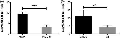
Figure 2. The expression of miR-34b was negatively associated with the expression of TGF-β1.
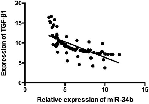
Figure 3. MiR-34b was decreased in cervical cancer tissues and inhibited the cell viability. (A) MiR-34b was significantly decreased in cervical cancer cell line compared with that in the corresponding normal cell line. (B) MiR-214 significantly inhibited the cell viability in day 2 and 3. **P < .01; ***P < .001 compared with the control group.
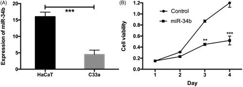
Figure 4. Over-expression of miR-34b would promote apoptosis. Representative Annexin V/PI staining of apoptosis cells in miR-34b or control miRNAs with ARPE-19 cells for 24 h.
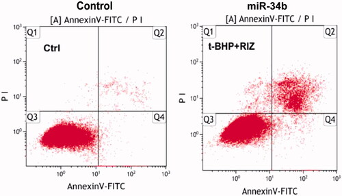
Figure 5. Effect of miR-34b on the migration of C33a cell line.
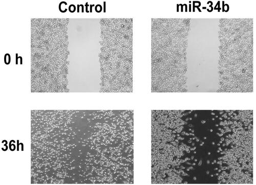
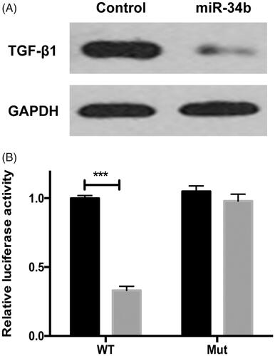
Figure 6. TGF-β1 was a direct target of miR-34b. (A) Protein level of TGF-β1 and GAPDH was detected by Western blot in C33a cells transfected with miR-34b/ctrl. (B) C33a cells were co-transfected with miR-34a and WT or Mut 3’-UTR luciferase reporter construct. ***P < .001 compared with the control group.
Availability of data and materials
The datasets used and/or analyzed during the present study are available from the corresponding author on reasonable request.