Figures & data
Figure 1. Determination of HBx expression in hepatocytes. HBx-encoding plasmids were transfected into L-02 hepatocytes for 48 h. Western blot analysis revealed the overexpression of HBx in normal human L-02 hepatocytes.
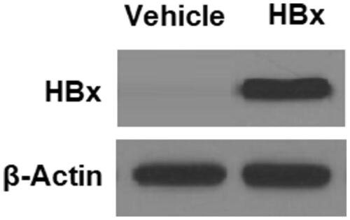
Figure 2. HBx reduces the expression of orexin-1 receptor (OX1R) in L-02 hepatocytes. The HBx-encoding plasmids were transfected into L-02 hepatocytes for 48 h. (A) Real-time PCR analysis revealed that overexpression of HBx decreased the expression of OX1R at the mRNA level; (B) Western blot analysis revealed that overexpression of HBx reduced the expression of OX1R at the protein level (*p < .01 vs vehicle group).
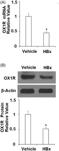
Figure 3. Orexin A mitigates HBx-induced oxidative stress in L-02 hepatocytes. L-02 hepatocytes were transfected with the HBx-encoding plasmid for 24 h, followed by treatment with orexin A at a concentration of 100 or 300 nM for another 24 h. (A) 4-HNE expression was measured by the immunostaining; (B) ROS was measured by the DCFH-DA assay (*, #, $, p < .01 vs previous column group).
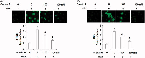
Figure 4. Orexin A ameliorates HBx-induced expression of NOX-4 in normal human L-02 hepatocytes. L-02 hepatocytes were transfected with the HBx-encoding plasmid for 24 h, followed by treatment with orexin A at a concentration of 100 or 300 nM for another 24 h. (A) Expression of NOX-4 at the mRNA levels determined by the real time analysis; (B). Expression of NOX-4 at the protein levels determined by the western blot analysis (*, #, $, p < .01 vs previous column group).
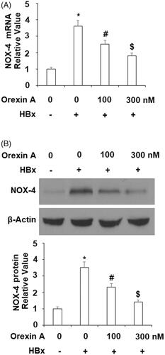
Figure 5. Orexin A ameliorates HBx-induced mitochondrial dysfunction in normal human L-02 hepatocytes. L-02 hepatocytes were transfected with the HBx-encoding plasmid for 24 h, followed by treatment with orexin A at a concentration of 100 or 300 nM for another 24 h. (A) Mitochondrial membrane potential (MMP) was measured by TMRM; (B) Intracellular ATP levels (*, #, $, p < .01 vs previous column group).
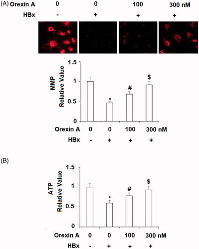
Figure 6. Orexin A inhibits HBx-induced expression and secretion of pro-inflammatory cytokines in normal human L-02 hepatocytes. L-02 hepatocytes were transfected with the HBx-encoding plasmid for 24 h, followed by treatment with orexin A at a concentration of 100 or 300 nM for another 24 h. (A) Expression of IL-8, TNF-α and CXCL2 at the mRNA levels was determined by real time PCR analysis; (B) Expression of IL-8, TNF-α and CXCL2 at the protein levels was determined by ELISA assay (*, #, $, p < .01 vs previous column group).
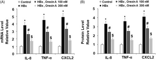
Figure 7. Orexin A attenuates HBx-induced cytotoxicity. L-02 hepatocytes were transfected with the HBx-encoding plasmid for 24 h, followed by treatment with orexin A at a concentration of 100 or 300 nM for another 24 h. Release of LDH was assayed (*, #, $, p < .01 vs previous column group).
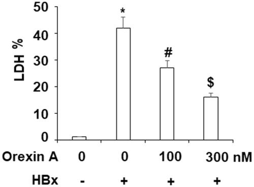
Figure 8. Orexin A suppresses HBx-induced phosphorylation of JNK in L-02 hepatocytes. L-02 normal hepatocytes were transfected with the HBx-encoding plasmid. After 24 h, cells were treated with orexin A at the concentrations of 100 and 300 nM for another 2 h. Phosphorylated and total levels of JNK were determined using western blot analysis (*, #, $, p < .01 vs previous column group).
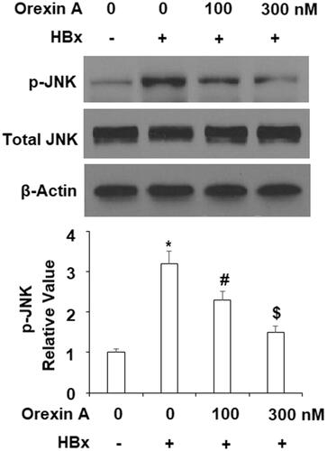
Figure 9. Orexin A treatment inhibited HBx-induced activation of NF-κB in L-02 hepatocytes. L-02 normal hepatocytes were transfected with HBx-encoding plasmids. After 24 h, cells were treated with orexin A at the concentrations of 100 and 300 nM for another 24 h. (A) Nuclear translocation of p65; (B) Luciferase activity of NF-κB (*, #, $, p < .01 vs previous column group).
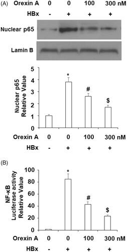
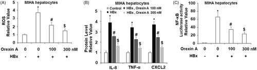
Figure 10. The protective effects of orexin A against HBx-induced cytotoxicity in human MIHA hepatocytes. Human MIHA hepatocytes were transfected with the HBx-encoding plasmid for 24 h, followed by treatment with orexin A at a concentration of 100 or 300 nM for another 24 h. (A) ROS was measured by DCFH-DA assay; (B). Secretions of IL-8, TNF-α and CXCL2 were measured by ELISA analysis; (C). Luciferase activity of NF-κB (*, #, $, p < 0.01 vs previous column group).