Figures & data
Figure 1. Ghrelin protects H9c2 cells against hypoxia/reoxygenation (H/R)-induced growth inhibition. (A) H9c2 cells were treated with various concentrations of ghrelin. (B) H9c2 cells were pre-treated with various concentrations of ghrelin and then subjected to H/R. Cell viability was tested by CCK-8 assay. (C–D) After the treatment of 0.1 μM ghrelin and stimulation with H/R, expression of cell-cycle-related proteins was measured by Western blot. *, ** and *** stand for p < .05, p < .01 and p < .001, respectively.
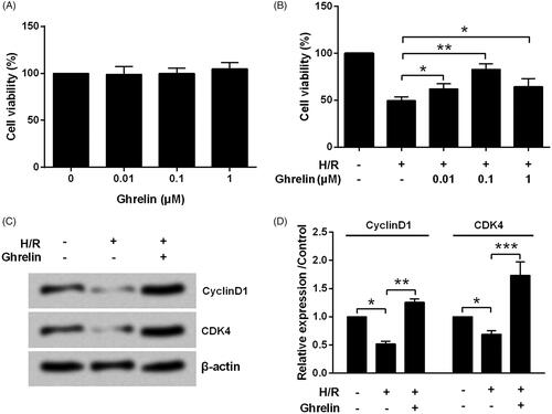
Figure 2. Ghrelin protects H9c2 cells against hypoxia/reoxygenation (H/R)-induced apoptosis and LDH release. H9c2 cells were pretreated with 0.1 μM ghrelin and were subjected to H/R. (A) Apoptosis rate, (B–C) expression of apoptosis-related proteins, and (D) the release of LDH were respectively assessed by flow cytometry, Western blot, and LDH Cytotoxicity Assay Kit. *, ** and *** stand for p < .05, p < .01 and p < .001, respectively.
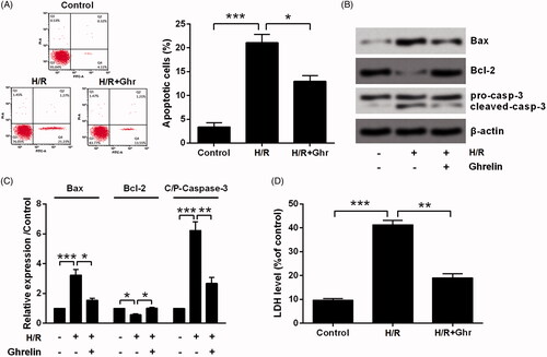
Figure 3. Ghrelin protects H9c2 cells against hypoxia/reoxygenation (H/R)-induced the expression of Cox-2 and iNOS. H9c2 cells were pretreated with 0.1 μM ghrelin and were subjected to H/R. Expression of Cox-2 and iNOS was measured by Western blot. *, ** and *** stand for p<.05, p<.01 and p<.001, respectively.
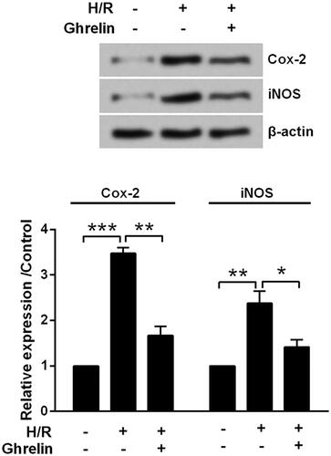
Figure 4. Ghrelin activates PI3K/AKT and AMPK pathways. H9c2 cells were pretreated with 0.1 μM ghrelin and were subjected to hypoxia/reoxygenation (H/R). Expression of proteins in (A) PI3K/AKT and (B) AMPK pathways was measured by Western blot. * and ** stand for p < .05 and p < .01, respectively.
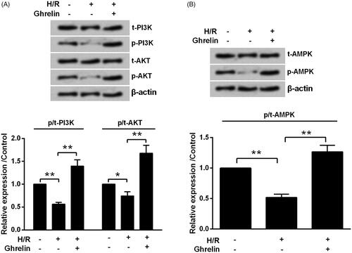
Figure 5. Ghrelin protects H9c2 cells against hypoxia/reoxygenation (H/R)-induced cell loss via PI3K/AKT and AMPK pathways. (A) Expression of proteins in PI3K/AKT and AMPK pathways was measured by Western blot, after the treatment of LY294002 and Compound C. H9c2 cells were pretreated with 0.1 μM ghrelin in the presence or absence of LY294002/Compound C and then subjected to H/R, (B) cell viability, (C) apoptosis rate, (D–E) expression of apoptosis-related proteins, and (F) the release of LDH were assessed by CCK-8 assay, flow cytometry, Western blot, and LDH Cytotoxicity Assay Kit. *, **, and *** stand for p < .05, p < .01 and p < .001, respectively.
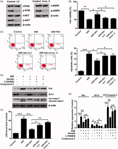
Figure 6. Ghrelin protects H9c2 cells against hypoxia/reoxygenation (H/R)-induced the expression of Cox-2 and iNOS via PI3K/AKT and AMPK pathways. H9c2 cells were pretreated with 0.1 μM ghrelin in the presence or absence of LY294002/Compound C and then were subjected to H/R. Expression of Cox-2 and iNOS was measured by Western blot. * and *** stand for p<.05 and p<.001, respectively.
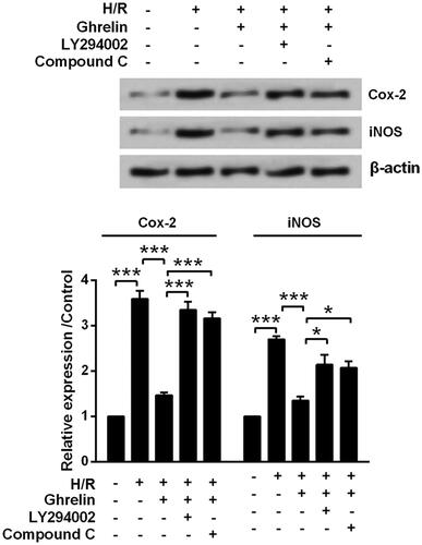
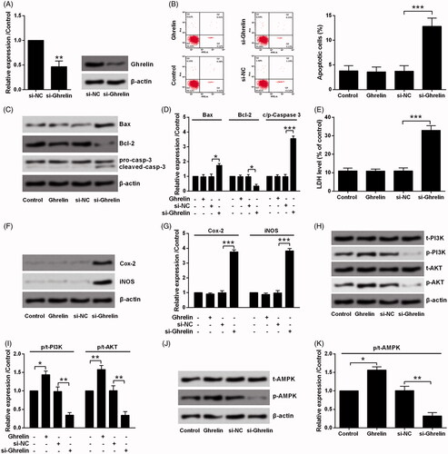
Figure 7. Silence of ghrelin induced H9c2 cell death via PI3K/AKT and AMPK pathways. (A) Expression of ghrelin in H9c2 cells was measured by qRT-PCR and Western blot, after transfection with the siRNA specific for ghrelin. H9c2 cells were treated with 0.1 μM ghrelin or transfected with ghrelin siRNA. (B) Apoptosis rate, (C–D) expression of apoptosis-related proteins, (E) the release of LDH, (F–G) expression of Cox-2 and iNOS, as well as the expression of proteins in (H–I) PI3K/AKT and (J–K) AMPK pathways were assessed by flow cytometry, Western blot, and LDH Cytotoxicity Assay Kit. *, ** and *** stand for p<.05, p<.01 and p<.001, respectively.