Figures & data
Figure 1. Lunasin ameliorated IL-1β-mediated reduction of cell proliferation. (A) Chondrocytes were incubated with lunasin (50, 100 and 500 μM) for 48 h. Cell proliferation was assessed by MTT. (B) Chondrocytes were cultured with IL-1β with or without lunasin (50, 100 μM). Chondrocytes proliferation was examined with MTT (*, #, p < .01).
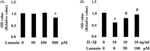
Figure 2. Lunasin ameliorated IL-1β-mediated induction of MMPs. Chondrocytes were stimulated with IL-1β or lunasin (50, 100 μM). (A) mRNA level of MMP-3, MMP-13. (B) Protein level of MMP-3, MMP-13 (*, #, p < .01).
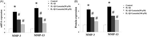
Figure 3. Lunasin ameliorated IL-1β-mediated decrease in TIMP-1 and TIMP-2. Chondrocytes were stimulated with IL-1β or lunasin (50, 100 μM). (A) mRNA level of TIMP-1, TIMP-2. (B) Protein level of TIMP-1, TIMP-2 (*, #, p < .01).
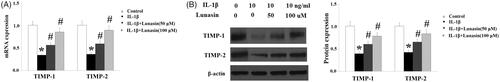
Figure 4. Lunasin mitigated IL-1β caused reduction of type II collagen. Chondrocytes were stimulated with IL-1β or lunasin (50, 100 μM). Col II, type II collagen. (A) Representative bands of type II collagen. (B) Quantitative analysis (*, #, p < .01).
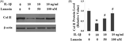
Figure 5. Lunasin abolished IL-1β-induced upregulation of IRF-1. Chondrocytes were stimulated with IL-1β or lunasin (50, 100 μM). (A) mRNA level of IRF-1. (B) Protein level of IRF-1 (*, #, p < .01).
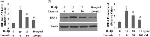
Figure 6. Lunasin suppressed IL-1β- induced activation of JAK2 and STAT1. Chondrocytes were stimulated with IL-1β or lunasin (50, 100 μM). (A) Representative bands of phosphorylated JAK2 and STAT1 at Ser 727, (B) Quantitative analysis (*, #, p < .01).
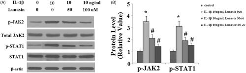
Figure 7. The JAK2/STAT1/IRF-1 pathway participates in the inhibitory effects of lunasin on MMPs. Chondrocytes were cultured with IL-1β and lunasin (100 μM) with or without the specific JAK2 activator coumermycin A1 (50 nM) or the specific JAK2 inhibitor AG490 (10 nM) for 24 h. (A) mRNA levels of MMPs. (B) Protein levels of MMPs (*, #, $, p < .01).
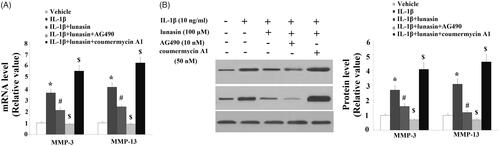
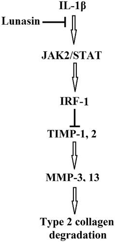
Figure 8. A graphical representation of the underlying mechanism.