Figures & data
Figure 1. miR-204 is low expressed in cervical cancer tissues and cells. (A) The expression of miR-204 in cervical cancer tissues; (B) the expression of miR-204 in cervical cancer cell lines. Compared to normal tissues or End/E6E7 cells, *p < .05.
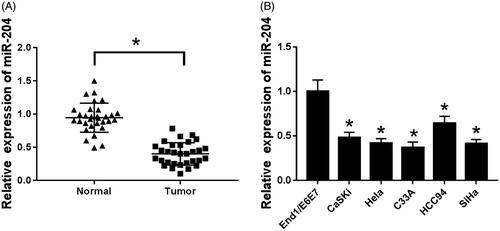
Figure 2. Overexpression of miR-204 inhibits proliferation of C33A cells. (A) The expression of miR-204 in C33A cells of each group; (B) cell viability of C33A cells in miR-204 transfection group. Compared with NC group and miR-con group, *p < .05.
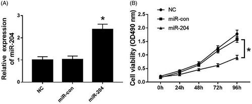
Figure 3. Overexpression of miR-204 inhibits autophagy and induces apoptosis in C33A cells. (A) The detection of apoptosis rate in each group; (B) the expression of apoptosis-related proteins Bcl-2, Bax, Caspase-3 and autophagy-related protein LC3I/II in each group. Compared with NC group and miR-con group, *p < .05.
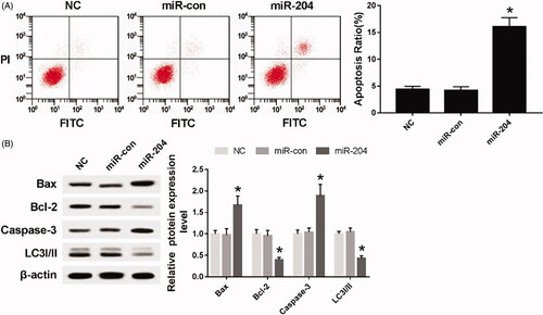
Figure 4. miR-204 targets and regulates ATF2 expression. (A) The binding site of miR-204 and ATF2 3′UTR; (B) the detection of double luciferase activity in each group; (C) the expression of ATF2 protein in each group. Compared with miR-con group, *p < .05; compared with anti-con group, #p < .05.
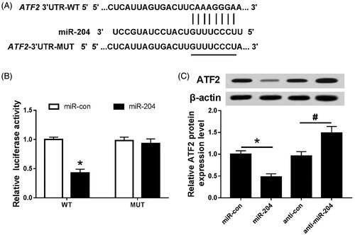
Figure 5. Effects of silencing ATF2 on proliferation, autophagy and apoptosis of C33A cells. (A) The expression of ATF2 and LC3I/II protein, (B) the cell viability of transfected cells and (C) the apoptosis rate of each group. Compared with NC group and si-con group, *p < .05.
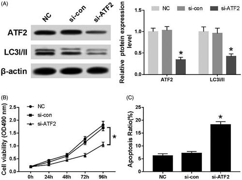
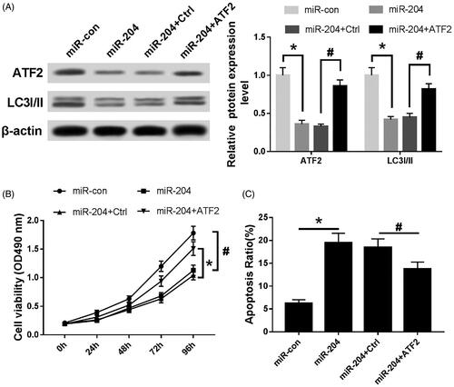
Figure 6. Effects of miR-204 and ATF2 on proliferation, autophagy and apoptosis of C33A cells. (A) The expression of ATF2 LC3I/II protein of each group, (B) MTT assay was utilized to detect cell viability and (C) the apoptosis rate of each group was evaluated by flow cytometry. Compared with miR-con group, *p < .05; compared with miR-204 + Ctrl group, #p < .05.