Figures & data
Figure 1. LPS stimulates HaCaT cell inflammation. (A) Cell viability, (B) cell apoptosis were detected by MMT assay and flow cytometry, respectively. (C,D) The apoptotic proteins cleaved-PARP, cleaved-caspase-3 and cleaved-caspase-9, (E) the accumulated levels of IL-1β and IL-6, (F, G) Cox-2 and iNOS were all examined by western blot. Results shown as mean ± SD and *p < .05 indicated significant difference.
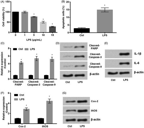
Figure 2. Emodin abated HaCaT cell inflammation induced by LPS. (A,B) Cell viability and (C) cell apoptosis were accessed by MTT assay and flow cytometry, respectively. (D,E) The accumulated levels of apoptosis related proteins cleaved-PARP, cleaved-caspase-3 and cleaved-caspase-9, (F) the expression of IL-1β and IL-6 and (G,H) Cox-2 and iNOS were detected by western blot.
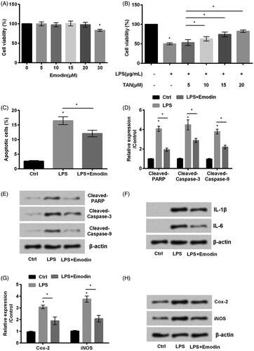
Figure 3. Emodin blocked NF-κB while activated PTEN/PI3K/AKT pathways in HaCaT cells. (A–D) The phosphorylation of NF-κB and PTEN/PI3K/AKT pathways related factors were detected by western blot. *p < .05.
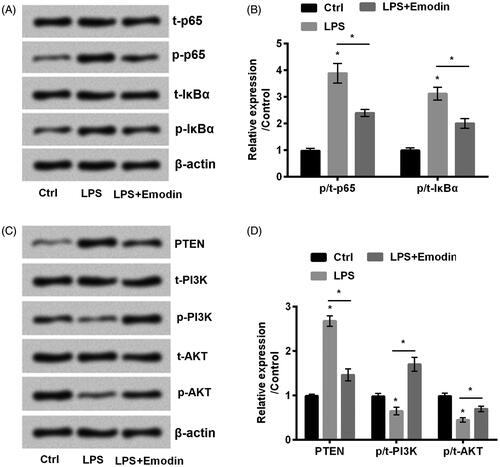
Figure 4. miR-21 expression was positively regulated by emodin. qRT-PCR was used in determining miR-21 expression in HaCaT cells. Results were shown as mean ± SD and *p < .05 indicated significant difference.
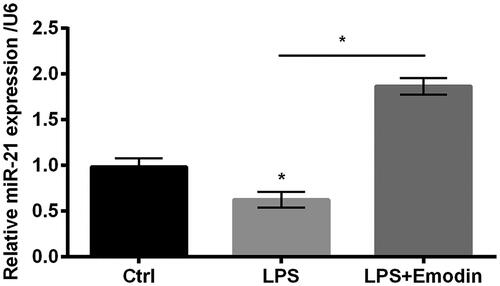
Figure 5. miR-21 downregulation aggravated LPS-induced inflammation in HaCaT cells. (A) miR-21 inhibitor was transduced into HaCaT cells. miR-21 level was evaluated by qRT-PCR. (B) Cell viability and (C) cell apoptosis were detected by MTT assay and flow cytometry, respectively. (D,E) The expression of apoptosis associated factors cleaved-PARP, cleaved-caspase-3 and cleaved-caspase-9, (F) the accumulated levels of inflammatory factors IL-1β and IL-6 and (G,H) Cox-2 and iNOS were detected by western blot. Results were shown as mean ± SD and *p < .05 indicated significant difference.
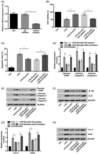
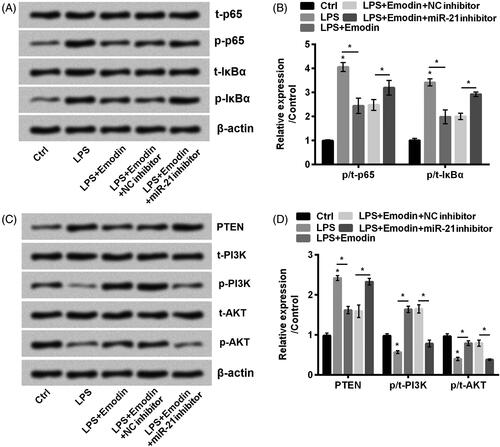
Figure 6. miR-21 downregulation promoted NF-κB while inactivated PTEN/PI3K/AKT pathways in HaCaT cells. (A–D) The phosphorylation of NF-κB and PTEN/PI3K/AKT pathways related factors were detected by western blot. Results were shown as mean ± SD and *p< .05 indicated significant difference.