Figures & data
Figure 1. BMP9 suppresses the protein expression of ALP, Runx2 and FAK of SMSCs. (A) The protein expression of ALP, Runx2 and FAK were detected via Western blot following different concentrations of BMP9. (B) Statistical analysis showed that 10–5 mol/l of BMP9 increased the highest expression of ALP and Runx2, exhibiting the best osteogenic capacity of SMSCs. *p < .05, **p < .01, as compared with 0 mol/l of BMP9.
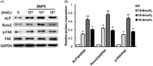
Figure 2. siRNA-induced knockdown decreases FAK expression of SMSCs. (A) Expression of FAK was observed via immunofluorescence assay following siRNA-induced knockdown. (B) Statistical analysis showed that siRNA-induced knockdown significantly inhibited fluorescence intensity of FAK. (C) siRNA-induced knockdown effectively suppressed the FAK expression following Western blot detection. **p < .01, as compared with BMP9-siFAK.
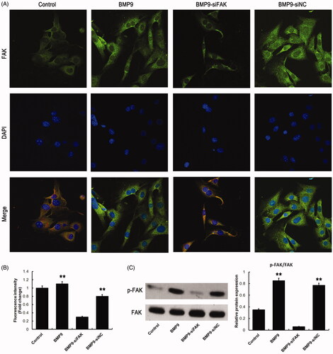
Figure 3. FAK knockdown inhibits BMP9-induced cell proliferation and migration of SMSCs in vitro. (A) Cell viability was markedly decreased by FAK knockdown following CCK8 assay. (B) GLUT3 expression was effectively suppressed by FAK knockdown following Western blot. (C) Cell proliferation was obviously decreased via FAK knockdown following EdU/ Hoechst staining. (D) Cell migration was significantly inhibited through FAK knockdown following transwell assay. **p < .01, as compared with BMP9-siFAK.
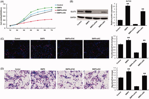
Figure 4. FAK knockdown decreases BMP9-induced osteogenesis of SMSCs in vitro. (A) BMP9-stimulated SMSCs were observed via ALP and alizarin red staining. (B) ALP absorption was obviously decreased via FAK knockdown. (C) Absorption of calcium deposition was effectively suppressed by FAK knockdown. (D) The protein expressions of ALP, Runx2, OCN and OPN were significantly inhibited through FAK knockdown following Western blot detection. *p < .05, **p < .01, as compared with BMP9-siFAK.
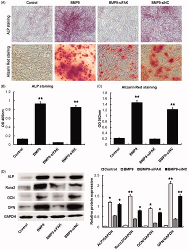
Figure 5. FAK knockdown inhibits BMP9-stimulated Wnt and MAPK pathway of SMSCs in vitro. (A) The expressions of Wnt related proteins, such as Wnt1, Wnt3a, β-catenin and p-GSK3β/GSK3β were significantly inhibited through FAK knockdown following Western blot detection. (B) The expressions of MAPK related proteins, such as p-JNK/JNK, p-ERK1/2/ERK1/2 and p-p38/p38 were markedly inhibited through FAK knockdown following Western blot detection. *p < .05, **p < .01, as compared with BMP9-siFAK.
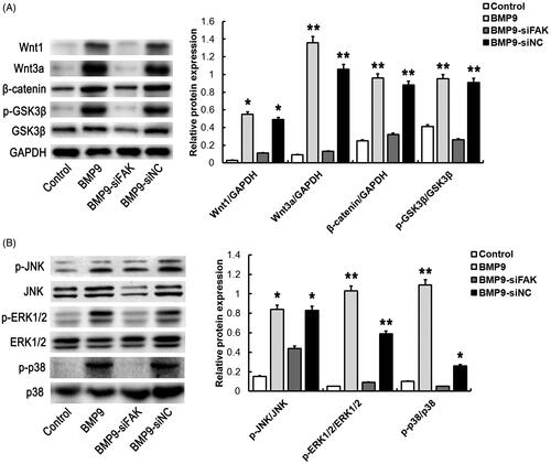
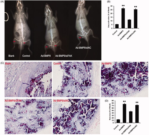
Figure 6. FAK knockdown inhibits BMP9-increased bone formation of SMSCs in vivo. (A) Cells-scaffold compound was observed via X-ray detection. (B) Statistical analysis indicated that FAK knockdown effectively suppressed BMP9-increased gray scale value. (C) Cells-scaffold compound was observed via HE staining. (D) Statistical analysis indicated that FAK knockdown markedly decreased BMP9-increased bone mass. **p < .01, as compared with Ad-BMP9/siFAK.