Figures & data
Figure 1. CCND1 expression in cervical carcinoma tissues and cell lines. (A) CCND1 expression in cervical carcinoma tissues and normal cervical tissues in different grades. (B) CCND1 expression in paired cervical carcinoma tissues and normal tissues. (C) CCND1 expression in cervical carcinoma cell lines and normal HUCECs. *p < .05 vs control.
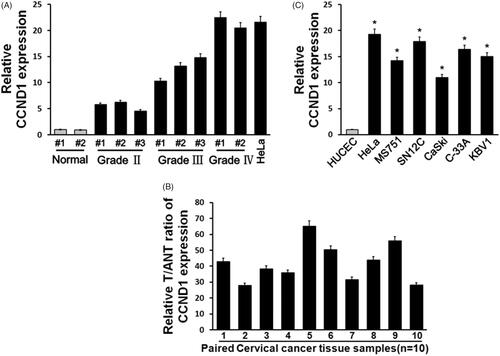
Figure 2. Baicalein reduced proliferation and apoptosis-related gene expression upregulated by CCND1. (A) CCND1 expression in HeLa cells after CCND1 or CCND1 siRNA transfection. (B) The results of cell cycle detection by flow cytometry. (C) HeLa cell proliferation detected using MTT assay. (D) Cell colony formation after CCND1 transfection or baicalein treatment. *p < .05 vs control.
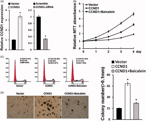
Figure 3. Baicalein reduced proliferation and apoptosis-related gene expression upregulated by CCND1. (A) Western blot was used to detect the expression of c-Myc and pRb in Hela cells. (B) Real-time PCR detection of apoptosis-related gene expression in HeLa cells. (C) Real-time PCR detection of stemness-related gene expressions in HeLa cells. *p < .05 vs control.
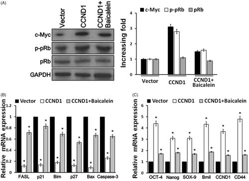
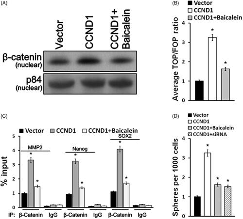
Figure 4. Baicalein targeted CCND1 via Wnt signaling pathway. (A) The expression of β-catenin in nucleus was detected using Western blotting. (B) TOP/FOP ratio of Wnt signaling pathway activity in HeLA cells was measured. (C) Sox 2, Nanog and MMP-2 levels were detected in HeLA cells. (D) Cells treated with baicalein or transfected with CCND1/CCND1 siRNA formed colonies. *p < .05 vs control.