
Figures & data
Figure 1. LPS prompted inflammation impairment in HK-2 cells. (A–C) The cell viability, cycle-related proteins expression and the apoptotic rate were detected by CCK-8, western blot and Annexin V-FITC/PI kit combined with flow cytometry. (D) qRT-PCR showed that the inflammatory factors mRNA expression levels were promoted by LPS. (E–H) The inflammatory factors were determined by ELISA. LPS: lipopolysaccharide; CCK-8: cell counting kit-8; FITC: fluorescein isothiocyanate; PI: propidium iodide; qRT-PCR: quantitative real-time polymerase chain reaction; mRNA: messenger RNA; ELISA: enzyme-linked immunosorbent assay. **p < .01, ***p < .001 compared to the corresponding group.
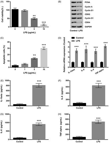
Figure 2. LPS led to miR-103 overproduction. The expression level of miR-103 was elevated by LPS. LPS: lipopolysaccharide; miR-103: microRNA-103. **p < .01 compared to the corresponding group.

Figure 3. miR-103 expression level was altered by miR-103 mimic and miR-103 inhibitor. Outcomes showed that miR-103 overexpressed after the transfection of miR-103 mimic, and miR-103 was decreased after the transfection of miR-103 inhibitor. miR-103 microRNA-103; **p < .01, ***p < .001 compared to the corresponding group.
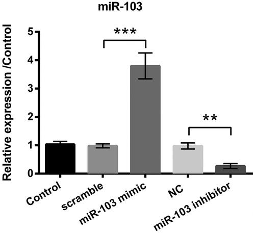
Figure 4. Influence of miR-103 on LPS-induced inflammatory injury. miR-103 mimic, miR-103 inhibitor and their negative controls (scramble and NC) were transfected into cells. (A–C) The cell viability, apoptotic rate and the apoptosis-related proteins expression were measured through CCK-8, and Annexin V-FITC/PI kit combined with flow cytometry and western blot, in proper sequence. (D) The inflammatory factors mRNA expression levels were tested by qRT-PCR. (E–H) Inflammatory factors production was determined by ELISA. LPS: lipopolysaccharide; miR-103: microRNA-103; NC: negative control; mRNA: messenger RNA; qRT-PCR: quantitative real-time polymerase chain reaction; CCK-8: cell counting kit-8; FITC: fluorescein isothiocyanate; ELISA: enzyme-linked immunosorbent assay. *p < .05, **p < .01, ***p < .001 in contrast with the corresponding group.
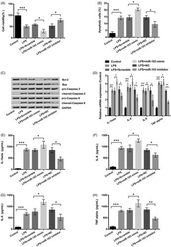
Figure 5. The c-Myc was a target of miR-103. The effect of miR-103 on c-Myc expression was detected by (A) qRT-PCR at the RNA level and (B) western blot at the protein level. (C) The luciferase activity was measured by luciferase reporter assay after the co-transfection of c-Myc-wt or c-Myc-mt and the miR-103 mimic. miR-103: microRNA-103; qRT-PCR: quantitative real-time polymerase chain reaction; c-Myc-wt: c-Myc-wild type; c-Myc-mt: c-Myc-mutant. *p < .05, **p < .01 in contrast with the corresponding group.
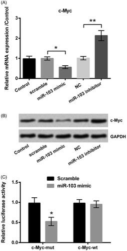
Figure 6. The impact of c-Myc on the protective effect of miR-103 inhibitor. After the respective transfection of miR-103 inhibitor and its negative control (NC) as well as the co-transfection of miR-103 inhibitor and si-c-Myc before LPS treatment, (A) the cell viability, (B) the cycle-related proteins expression, (C) the apoptosis rate, (D) the apoptosis-related proteins, (E) the inflammatory factors mRNA expression level and (F–I) the inflammatory factors real production were assessed. miR-103: microRNA-103; LPS: lipopolysaccharide; mRNA: messenger RNA. *p < .05, **p < .01, ***p < .001 in contrast with the corresponding group.
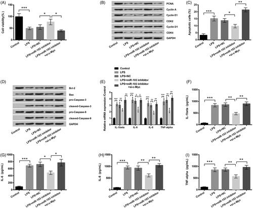
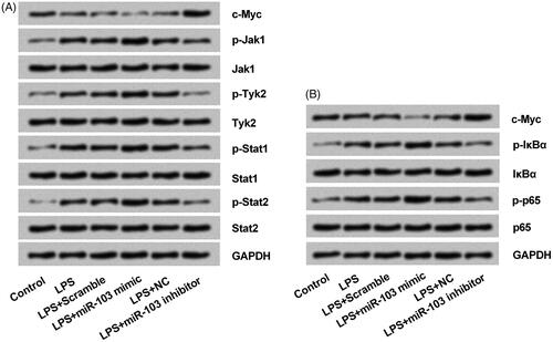
Figure 7. miR-103 regulated the NF-κB and JAK/STAT pathways activity through c-Myc modulation. (A) The expression of phosphorylated key proteins of NF-κB pathway and (B) JAK-STAT pathway were appraised by western blot. NF-κB: nuclear factor κB; JAK: janus kinase; STAT: signal transducers and activators of transcription; miR-103: microRNA-103.
Data availability
The datasets used and/or analysed during the current study are available from the corresponding author on reasonable request.