Figures & data
Figure 1. (A) The synthesis of AgNPs from aqueous leaf extract of Salvia miltiorrhiz was confirmed by changes in solution color from light yellow to dark brown. (B). UV-visible absorption spectrum of synthesized AgNPs.
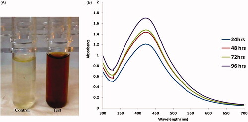
Figure 2. Dynamic light scattering (DLS) images of silver nanoparticles synthesized from Salvia miltiorrhiza and the size of the nanoparticles is 128 nm.
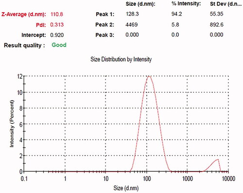
Figure 3. (A) Transmission electron microscopy (TEM) and (B) energy dispersive X-ray (EDX) and analysis silver nanoparticles synthesized from Salvia miltiorrhiza.
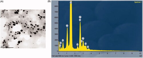
Figure 4. XRD analysis of silver nanoparticles (AgNPs) synthesized from Salvia miltiorrhiza.
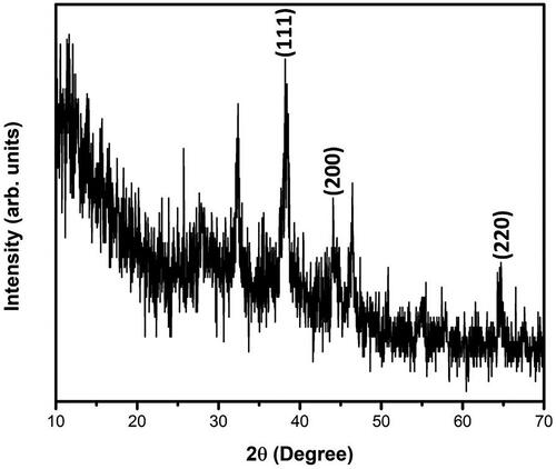
Figure 5. Fourier transforms infrared (FTIR) spectroscopy analysis of silver nanoparticles (AgNPs) synthesized from Salvia miltiorrhiza.
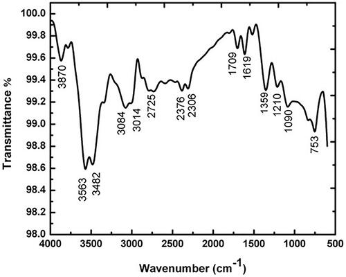
Figure 6. Images of antibacterial activities of silver nanoparticles against Salmonella typhi, Shigella flexneri, Streptococcus pyogene and Pseudomonas aeruginosa. A – 10 µg; B– 20 µg; C – 40 µg; D – 60 µg; E – positive control.

Table 1. Antimicrobial activity of AgNPs from salvia miltiorrhiza.
Figure 7. (A) Cytotoxic potential of AgNPs from Salvia miltiorrhiza in prostate cancer LNcap cell. (B). Effect of AgNPs from Salvia miltiorrhiza mediated ROS measurements in LNcap cells.
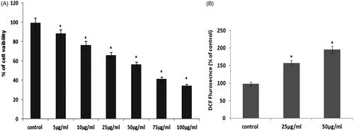
Figure 8. (A) AgNPs mediated apoptotic morphological studied by AO/PI staining. (B) AgNPs mediated nuclear fragmentation in LNcap cells studied by TUNEL assay. (C) DAPI staining analysis.
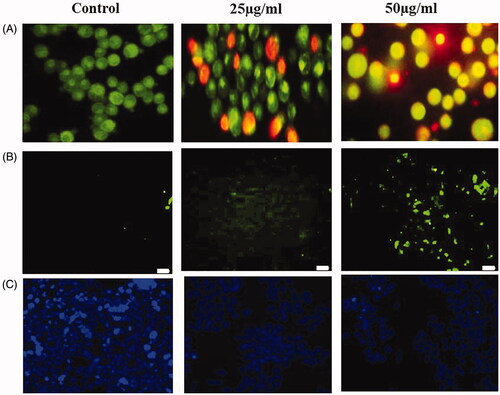
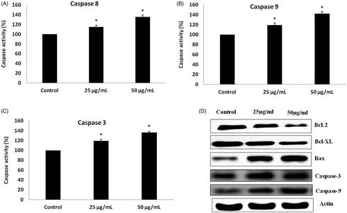
Figure 9. (A–C) Colorimetric assay for caspase-8, 9 & 3 activity expression in AgNPs treated LNcap cells. (D). Western blotting images for Bax, Bcl-xl, Bcl-2 and caspase-3 protein expression in AgNPs treated LNcap cells.