Figures & data
Figure 1. Hypoxia induced injury in H9c2 cells. (A) Cell viability, (B) cell migration; (C) cell invasion; (D) cell apoptosis and the expression of apoptosis-related proteins. The experiments were repeated three times. (E) Hypoxia promoted the expression of RP4 in H9c2 cells. The experiment was repeated three times. Data are expressed as mean ± SD. **p < .01 compared to control.
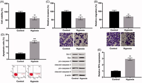
Figure 2. Overexpression of RP4 aggravated hypoxia-induced injury in H9c2 cells, while suppression of RP4 relieved the injury. (A) The expression of RP4 in H9c2 cells after transfection with pc-RP4, sh-RP4 and their NC. (B–E) H9c2 cells were transfected with pc-RP4, sh-RP4 and their NC under hypoxia condition. (B) Cell viability of different treatment groups; (C) cell migration of different treatment groups; (D) cell invasion of different treatment groups; (E) cell apoptosis and the expression of apoptosis-related proteins in different treatment groups. The experiments were repeated three times. Data are expressed as mean ± SD. *p < .05; **p < .01 and ***p < .001 compared to control.
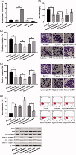
Figure 3. RP4 negatively regulated the expression of miR-939 in H9c2 cells, and overexpression of miR-939 abrogated the effects of overexpression of RP4 on hypoxia-induced injury. (A) The expression of miR-939 in H9c2 cells after transfection with pc-RP4, sh-RP4 and their NC. (B) The expression of miR-939 in H9c2 cells after transfection with miR-939 mimic, miR-939 inhibitor and their NC. (C–F) H9c2 cells were co-transfected with pc-RP4, miR-939 mimic and/or their respective NC under hypoxia condition. (C) Cell viability of different treatment groups; (D) cell migration of different treatment groups; (E) cell invasion of different treatment groups; (F) cell apoptosis and the expression of apoptosis-related proteins in different treatment groups. The experiments were repeated three times. Data are expressed as mean ± SD. *p < .05; **p < .01 and ***p < .001 compared to control.
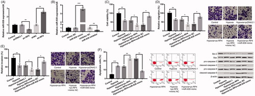
Figure 4. Bnip3 was a target of miR-939, and miR-939 negatively regulated Bnip3 expression. (A) The binding sequence between miR-939 and Bnip3. (B) Luciferase reporter assay revealed the target relationship between miR-939 and Bnip3. (C–D) The mRNA and protein expression of Bnip3 in H9c2 cells after transfection with miR-939 mimic, miR-939 inhibitor and their NC. The experiments were repeated three times. Data are expressed as mean ± SD. *p < .05 and **p < .01 compared to control.
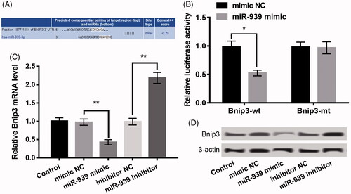
Figure 5. miR-939 prevented hypoxia-induced injury in H9c2 cells by targeting Bnip3. (A) Bnip3 expression in H9c2 cells after transfection with pEX-Bnip3, si-Bnip3 and their NC. (B–E) H9c2 cells were co-transfected with pc-RP4, miR-939 mimic, pEX-Bnip3 and/or their respective NC under hypoxia condition. (B) Cell viability of different treatment groups; (C) cell migration of different treatment groups; (D) cell invasion of different treatment groups; (E) cell apoptosis and the expression of apoptosis-related proteins in different treatment groups. The experiments were repeated three times. (F) The expression of Wnt/β-catenin pathway-related proteins in H9c2 cells that were co-transfected with pc-RP4, miR-939 mimic, pEX-Bnip3 and/or their respective NC under hypoxia condition. The experiments were repeated three times. Data are expressed as mean ± SD. *p < .05; **p < .01 and ***p < .001 compared to control.
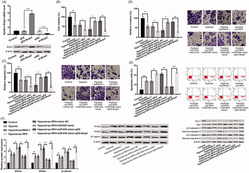
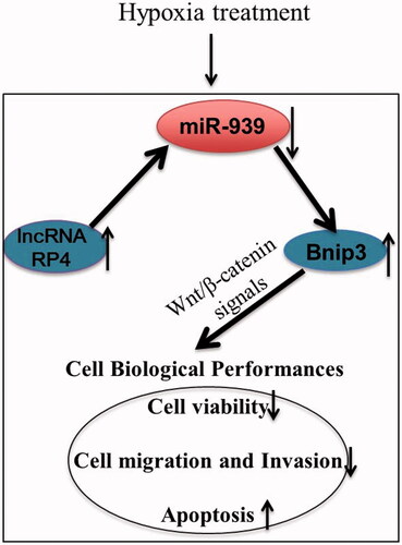
Figure 6. The regulatory mechanism of lncRNA RP4 in hypoxia induced injury in H9c2 cells.