Figures & data
Table 1. Demographic characteristics and clinical features of lung cancer patients.
Figure 1. LncRNA SDPR-AS was down-regulated NSCLC tissues (A) and cells (B). NSCLC: non-small cell lung cancer AC: adenocarcinoma; SCC: squamous cell carcinoma; LCC: large cell carcinoma; SCLC: small cell lung cancer. Data were presented as mean ± standard deviation. *p < .05, **p < .01 and ***p < .01 compared with corresponding controls.
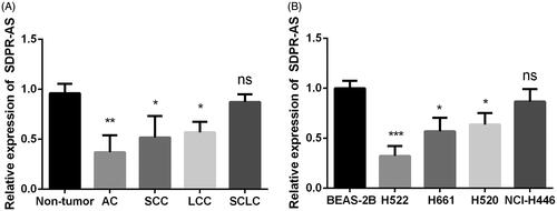
Figure 2. Down-regulation of SDPR-AS promoted NSCLC cell proliferation and inhibited cell apoptosis. NSCLC cell lines H522, H661 and H520 were transfected with pcDNA-SDPR-AS, sh-SDPR-AS or their corresponding controls. (A) SDPR-AS was confirmed to be successfully overexpressed and knocked down in H522, H661 and H520 cells. (B) Effects of SDPR-AS dysregulation on H522 cell viability, colony forming ability and apoptosis. (C) Effects of SDPR-AS dysregulation on H661 cell viability, colony forming ability and apoptosis. (D) Effects of SDPR-AS dysregulation on H520 cell viability, colony forming ability and apoptosis. Data were presented as mean ± standard deviation. *p<.05, **p<.01 and ***p<.01 compared with corresponding controls.
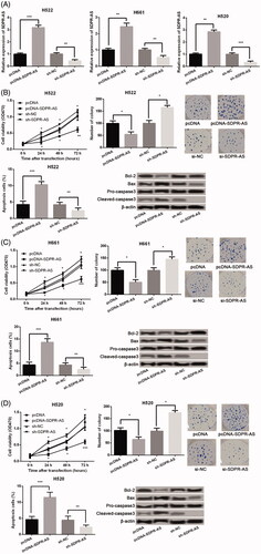
Figure 3. Down-regulation of SDPR-AS promoted the migration and invasion of H522 (A), H661 (B) and H520 (C) cells. Data were presented as mean ± standard deviation. *p < .05, **p < .01 and ***p < .01 compared with corresponding controls.
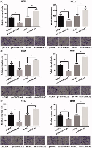
Figure 4. The expression of SDPR was positively regulated by SDPR-AS. (A) The expression of SDPR in AC, SCC, LCC, SCLC and non-tumor tissues. (B) The expression of SDPR in the human AC cell line H522, LCC cell line H661, SCC cell line H520, SCLC cell line NCI-H446 and normal lung epithelial cell line BEAS-2B. (C) The expression of SDPR in NSCLC cell lines H522, H661 and H520 that were transfected with pcDNA-SDPR-AS, sh-SDPR-AS or their corresponding controls. Data were presented as mean ± standard deviation. *p < .05, **p < .01 and ***p < .01 compared with corresponding controls.
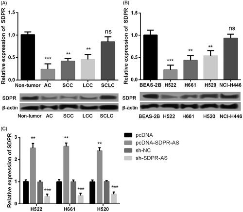
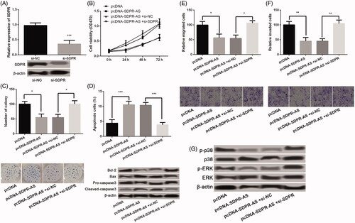
Figure 5. Effects of SDPR-AS on NSCLC cells were through regulation of SDPR. (A) The transfection efficiency of SDPR knockdown in H522 cells that were transfected with si-SDPR and si-NC. (B–F) The combined effects of SDPR-AS overexpression and SDPR knockdown on H522 cell viability, colony forming ability, apoptosis, migration and invasion. (G) The expression of p38 MAPK/ERK signaling pathway-related proteins in H522 cells that were transfected with pcDNA-SDPR-AS, pcDNA, si-NC and/or si-SDPR. Data were presented as mean ± standard deviation. *p < .05, **p < .01 and ***p < .01 compared with corresponding controls.