
Figures & data
Figure 1. Chemical construction of luteolin.
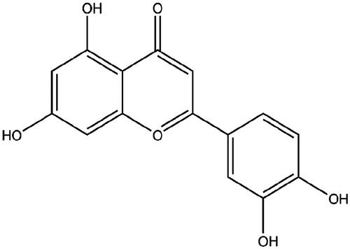
Figure 2. Luteolin suppressed breast cancer cells growth and EMT progress. MCF-7 and MDA-MB-453 cells were administrated with 10 μM luteolin for 24. (A) Cell viability, (B) apoptosis rate, (C–E) accumulation of apoptosis-related proteins, and (F–H) expression of EMT-markers were checked by CCK-8 assay, FITC-PI double-staining and Western blot. 5 ng/mL TGFβ1 was utilized to treat cells for 24 h to make an experimental EMT model. *, ** and *** stand for p values <.05, .01, and .001.
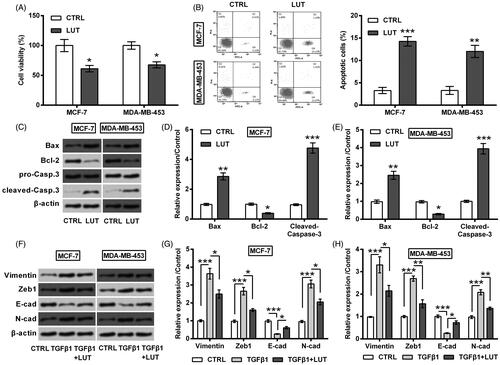
Figure 3. Luteolin increased miR-203 expression. (A) miR-203 expression in 22 pair of breast cancer tissues and the matched paracancerous tissues was monitored by qRT-PCR. (B) miR-203 expression in MCF-7 and MDA-MB-453 cells was monitored by qRT-PCR after treating with luteolin. **and ***stand for p values <.01 and .001.
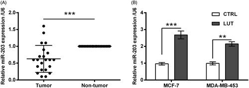
Figure 4. Luteolin suppressed breast cancer cells growth via miR-203. (A) miR-203 expression in MCF-7 and MDA-MB-453 cells was monitored by qRT-PCR after transfection with miR-203 inhibitor and NC. The transfected cells were treated by luteolin. (B) Cell viability, (C) apoptosis rate, and (D–F) accumulation of apoptosis-related proteins were checked by CCK-8 assay, FITC-PI double-staining and Western blot. *, ** and *** stand for p values <.05, .01 and .001.
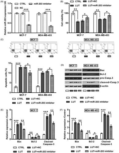
Figure 5. Luteolin suppressed breast cancer cells EMT progress via miR-203. MCF-7 and MDA-MB-453 cells were transfected with miR-203 inhibitor and NC. The transfected cells were treated by luteolin. (A–C) Expression of EMT-markers was checked by Western blot. 5 ng/mL TGFβ1 was utilized to treat cells for 24 h to make an experimental EMT model. *, ** and *** stand for p values <.05, .01 and .001.
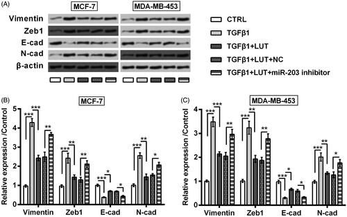
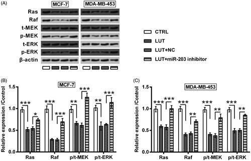
Figure 6. Luteolin inhibited Ras/Raf/MEK/ERK signalling via miR-203. MCF-7 and MDA-MB-453 cells were transfected with miR-203 inhibitor and NC. The transfected cells were treated by luteolin. The expression of Ras and Raf, as well as the phosphorylation of MEK and ERK were checked by Western blot. *, ** and *** stand for p values <.05, .01 and .001.
Data availability statement
The datasets used and/or analysed during this study are available from the corresponding author on reasonable request.