Figures & data
Table 1. The Association of miR-638 expression with clinical characteristics of OSCC patients.
Figure 1. Decreased expression of miR-638 in OSCC tissues (A) and cells (B). **P < .01; *P < .05.
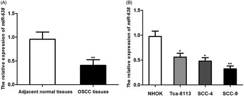
Figure 2. The transfection with miR-638 mimic could enhance the expression of miR-638, while transfection with miR-638 inhibitor down-regulated miR-638 (A). Enforced miR-638 could significantly suppress OSCC cells’ proliferation (B), migration (C) and invasion (D), and the knockdown of miR-638 promoted malignant behaviors of OSCC cells. **P < .01; *P < .05.
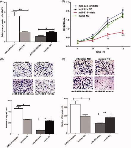
Figure 3. Bioinformatics analysis demonstrated that the 3′UTR of PLD1 gene had complementary sequences of miR-638 (A). In cells transfected by PLD1-wt, the presence of miR-638 mimic could significantly reduce luciferase activity of the cells, while the co-transfection with miR-638 and PLD1-mt had no obvious influences on luciferase activity of the cells (B). **P < .01.
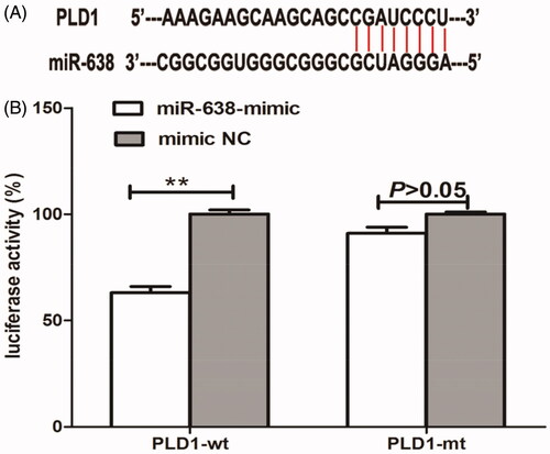
Figure 4. The expression of PLD1 was significantly up-regulated in SCC-9 cells, compared to non-cancerous HPK cell line (A). Moreover, enforced expression of miR-638 could obviously suppress the expression of PLD1, while miR-638 inhibition up-regulated PLD1 in OSCC (B). ***P < .001; **P < .01.
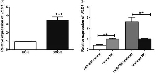
Figure 5. Western blot images for the expressions of wnt/β-catenin pathway-related proteins in transfected cells (A). Quantitative analysis for western blot results (B). **P < .01; *P < .05.
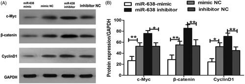
Figure 6. Western blot analysis for proteins in wnt/β-catenin pathway after the co-transfection with miR-638 inhibitor and si-PLD1 (A). Quantitative analysis for western blot results (B). **P < .01; *P < .05.
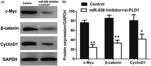
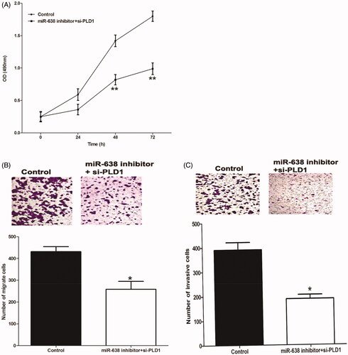
Figure 7. Compared to cells transfected by miR-638 inhibitor (control), the co-transfection with miR-638 inhibitor and si-PLD1 significantly suppressed cell proliferation (A), migration (B) and invasion (C). **P < .01; *P < .05.