Figures & data
Figure 1. Saxagliptin inhibited expression of MMP-1, MMP-3, MMP-13. Primary human chondrocytes were treated with 100 μg/ml AGEs in the presence or absence of 0.5 and 1 μM saxagliptin for 24 h. (A) Gene expressions of MMP-1, MMP-3, and MMP-13 were determined by real-time PCR analysis; (B) Protein expressions of MMP-1, MMP-3, and MMP-13 was determined by ELISA (*, #, $, p < .01 vs. previous column group).
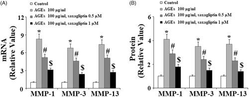
Figure 2. Saxagliptin inhibited degradation of type II collagen. Primary human chondrocytes were treated with 100 μg/ml AGEs in the presence or absence of 0.5 and 1 μM saxagliptin for 24 h. Type II collagen was determined by western blot analysis (*, #, $, p < .01 vs. previous column group).
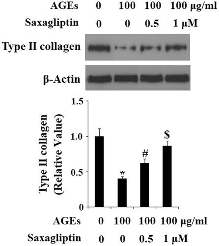
Figure 3. Saxagliptin inhibited expression of ADAMTS-4 and ADAMTS-5. Primary human chondrocytes were treated with 100 μg/ml AGEs in the presence or absence of 0.5 and 1 μM saxagliptin for 24 h. (A) mRNA expression of ADAMTS-4 and ADAMTS-5 determined by real-time PCR; (B) Protein expression of ADAMTS-4 and ADAMTS-5 determined by western blot analysis (*, #, $, p < .01 vs. previous column group).
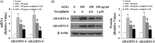
Figure 4. Saxagliptin inhibited aggrecan degradation. Primary human chondrocytes were treated with 100 μg/ml AGEs in the presence or absence of 0.5 and 1 μM saxagliptin for 24 h. Aggrecan was determined by western blot analysis (*, #, $, p < .01 vs. previous column group).
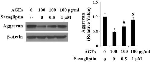
Figure 5. Saxagliptin inhibited oxidative stress. Primary human chondrocytes were treated with 100 μg/ml AGEs in the presence or absence of 0.5 and 1 μM saxagliptin for 24 h. (A) Reactive oxygen species (ROS) was determined by DCFH-DA; (B) GSH level (*, #, $, p < .01 vs. previous column group).
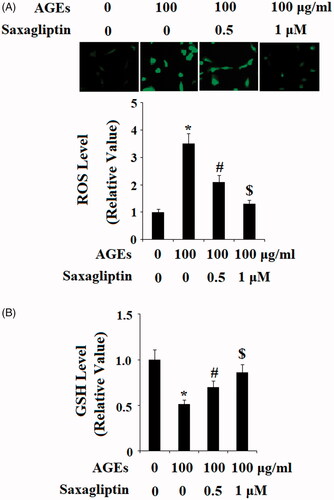
Figure 6. Saxagliptin inhibited phosphorylation of p38. Primary human chondrocytes were treated with 100 μg/ml AGEs in the presence or absence of 0.5 and 1 μM saxagliptin for 2 h. Phosphorylated and total p38 were determined by western blot analysis (*, #, $, p < .01 vs. previous column group).
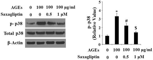
Figure 7. Saxagliptin inhibited phosphorylation and degradation of IκBα. Primary human chondrocytes were treated with 100 μg/ml AGEs in the presence or absence of 0.5 and 1 μM saxagliptin for 6 h. Phosphorylated and total IκBα were determined by western blot analysis (*, #, $, p < .01 vs. previous column group).
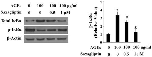
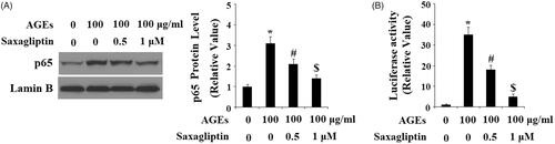
Figure 8. Saxagliptin inhibited activation of NF-κB. Primary human chondrocytes were treated with 100 μg/ml AGEs in the presence or absence of 0.5 and 1 μM saxagliptin for 24 h. (A) Nuclear level of p65; (B) Luciferase activity of p65 (*, #, $, p < .01 vs. previous column group).