Figures & data
Figure 1. Characterization of DQA modified paclitaxel plus ligustrazine micelles. Notes: (A) A schematic representative of DQA modified paclitaxel plus ligustrazine micelles. (B) TEM image of DQA modified paclitaxel plus ligustrazine micelles. (C) Zeta potential of DQA modified paclitaxel plus ligustrazine micelles. (D) Partical size of DQA modified paclitaxel plus ligustrazine micelles.
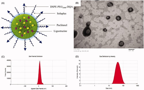
Table 1. Characterization of the varying micelles.
Figure 2. Cytotoxicity on A549 cells after treatments with the varying formulations. Notes: (A) Cytotoxicity of free drugs, I, vs. free ligustrazine; II, vs. free paclitaxel; III, vs. free paclitaxel: free ligustrazine =1:1; IV, vs. free paclitaxel: free ligustrazine = 1:3. (B) Cytotoxicity of the varying micellar formulations. 1, vs. Blank micelles; 2, vs. Paclitaxel micelles; 3, vs. DQA modified paclitaxel micelles; 4, vs. Paclitaxel plus ligustrazine micelles. Data are presented as mean ± SD (n = 3). p < .05.
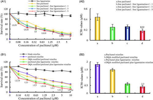
Figure 3. Cellular uptake and targeting effects after incubation with the varying formulations. Notes: (A) Cellular uptake of A549 cells. (B) Fluorescence intensity of coumarin 6 in A549 cells. a. Blank micelles; b. Coumarin micelles; c. DQA modified coumarin micelles; d. Coumarin plus ligustrazine micelles; e. DQA modified coumarin plus ligustrazine micelles. p < .05, 1, vs. a; 2, vs. b; 3, vs. c; 4, vs. d.
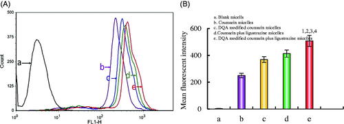
Figure 4. Destructive effects on VM channels in vitro after incubation with the varying formulations. Notes: a. Blank micelles; b. Paclitaxel micelles; c. DQA modified paclitaxel micelles; d. Paclitaxel plus ligustrazine micelles; e. DQA modified paclitaxel plus ligustrazine micelles; f. VM channel correlation index ratio. p < .05, 1 vs a, 2 vs b, 3 vs c, 4 vs d.
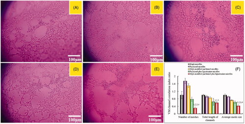
Figure 5. Inhibiting invasion on A549 cells in vitro after incubation with the varying formulations. Notes: a. Blank micelles; b. Paclitaxel micelles; c. DQA modified paclitaxel micelles; d. Paclitaxel plus ligustrazine micelles; e. DQA modified paclitaxel plus ligustrazine micelles; f. Cell invasion rate. p < .05, 1 vs a, 2 vs b, 3 vs c, 4 vs d.
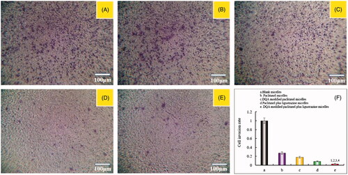
Figure 6. Blocking wound healing effects on A549 cells in vitro after incubation with the varying formulations. Notes: (A) Blocking wound healing effects; (B) Wound healing rate (%). a. Blank micelles; b. Paclitaxel micelles; c. DQA modified paclitaxel micelles; d. Paclitaxel plus ligustrazine micelles; e. DQA modified paclitaxel plus ligustrazine micelles; f. Cell invasion rate. p < .05, 1 vs Blank micelles, 2 vs Paclitaxel micelles, 3 vs DQA modified paclitaxel micelles, 4 vs Paclitaxel plus ligustrazine micelles.
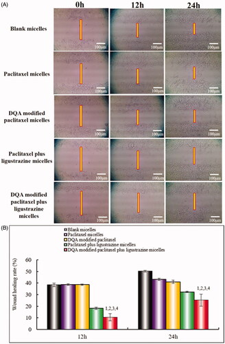
Figure 7. Adhesion rate of A549 cells on Matrigel after incubation with the varying formulations. Notes: (A) Cellular survival rate of A549 cells after incubation with the varying formulations for 12 h. (B) Adhesion rate of A549 cells on Matrigel. p < .05.
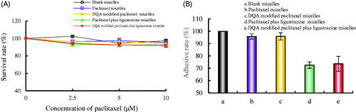
Figure 8. Regulating effects on metastasis-related proteins in A549 cells after treatments with the varying formulations. Notes: a. Blank micelles; b. Paclitaxel micelles; c. DQA modified paclitaxel micelles; d. Paclitaxel plus ligustrazine micelles; e. DQA modified paclitaxel plus ligustrazine micelles. p < .05, 1, vs. a; 2, vs. b; 3, vs. c. Data are presented as mean ± SD (n = 4).
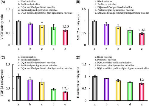
Figure 9. Fluorescence microscopy images of the fluorescence intensity of activated apoptotic enzymes Caspase-3, Caspase-8 and Caspase-9 proteins in A549 cells. Scale bar = 100 μm.
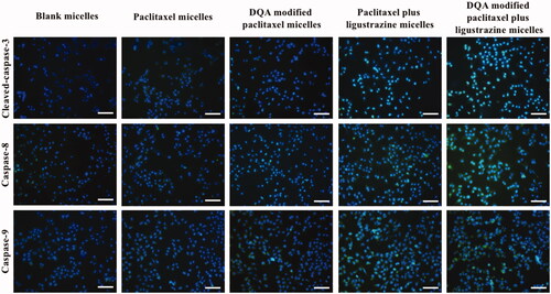
Figure 10. In vivo real-time imaging observation after intravenous administration of the varying formulations.
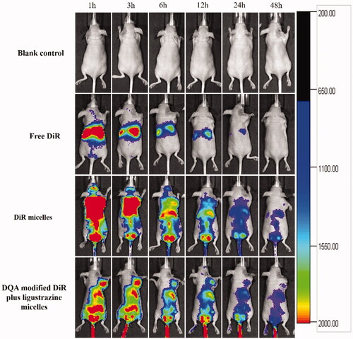
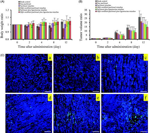
Figure 11. Anticancer effects in A549 cells xenografts mice after treatment with the varying formulations. Notes: (A) Body weight changes. (B) Tumour volume changes. (C) Representative images of tumour cells apoptosis were detected by TUNEL, Scale bar = 100 μm. Where a. blank control, b. free paclitaxel, c. paclitaxel micelles, d. DQA modified paclitaxel micelles, e. paclitaxel plus ligustrazine micelles and f. DQA modified paclitaxel plus ligustrazine micelles.