Figures & data
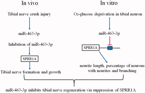
Figure 1. MiR-463-3p was first increased and then decreased after tibial nerve injury. The tibial nerve tissues were isolated and acquired in mice from lesions at post-injury 0, 3, 7, 12, 18 or 24 days, respectively. The tibial nerve cells from mice were primary cultured and received the OGD treatment. (A) The expression of miR-463-3p was analyzed by qRT-PCR in tibial nerve tissues. (B) The level of miR-463-3p was detected in tibial nerve cells at 0, 0.5, 1.0, 2.0 h under OGD. Data are shown as mean ± SD (N = 4). *p < .05 and #p < .05 vs. day 7 or 0.5 h.
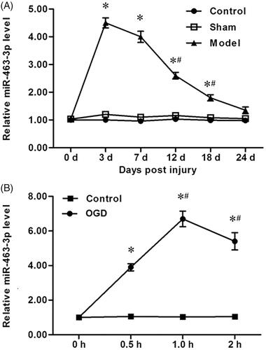
Figure 2. MiR-463-3p mimic inhibits neurite elongation and branching in vitro. The tibial nerve cells were transfected with miR-463-3p mimic or mimic-NC. (A) The level of miR-463-3p among groups was measured with qRT-PCR. (B) The length of neurites per cell was increased after miR-463-3p mimic transfection. (C) At 48 h, the percentage of cells with neurites was decreased in miR-463-3p mimic group relative to control group. (D) The number of branches per cell was increased in miR-463-3p mimic group. N = 4, *p<.05.
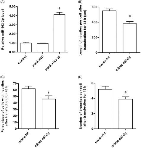
Figure 3. MiR-463-3p antagonist enhances neurite elongation and branching in vitro. The tibial nerve cells were transfected with miR-463-3p antagonist or anta-NC, respectively. (A) The level of miR-463-3p among groups. (B) The length of neurites per cell after transfection. (C) The percentage of cells with neurites among groups. (D) The number of branches per cell among groups. N = 4, *p<.05.
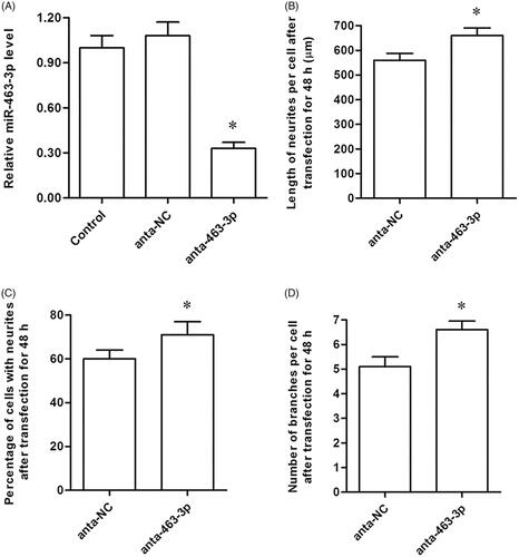
Figure 4. MiR-463-3p is direct target of SPRR1A. (A) Prediction result of potential binding sites between miR-463-3p and 3′UTR of SPRR1A by using the TargetScan. (B) Sequence of the wild-type and the mutant form 3′UTR of SPRR1A mRNA in mice contains a putative microRNA-463-3p-binding sites. (C) Relative luciferase activity in HEK-293T cells after transfection with miR-463-3p mimic and SPRR1A-3′UTR-WT or SPRR1A-3′UTR-MUT. Data are shown as mean ± SD (N = 4). *p<.05.
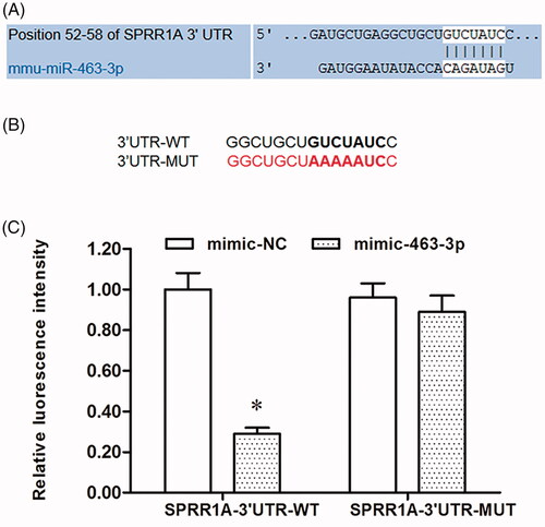
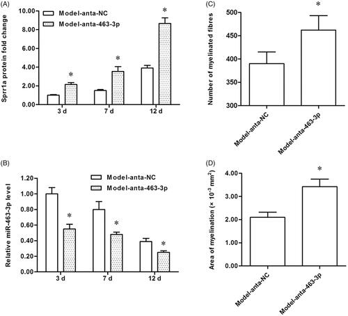
Figure 5. MiR-463-3p antagonist causes tibial nerve regeneration and promotes SPRR1A expression in vivo. The tibial nerve injury model of mice was established, miR-463-3p antagonist or anta-NC was subcutaneously injected into mice. The tibial nerve lesions tissues were isolated from mice at post-injury 3, 7 or 12 days, respectively. (A) The SPRR1A expression among groups were measured by using qRT-PCR. (B) The level of miR-463-3p was detected by qRT-PCR. Mice tibial nerve tissues were stained with toluidine blue to tibial nerve regeneration. (C) The total number of myelinated fibers was increased in anta-miR-463-3p group. (D) The area of myelination was increased after anta-miR-463-3p transfection. Data are shown as mean ± SD (N = 4). *p<.05.