Figures & data
Table 1. Primer sequences used in qRT-PCR experiments.
Figure 1. Juglanin reversed IL-1β-induced reduction in cell viability. (A) Cells were treated with 0, 10, 20, 40 and 80 μM of juglanin in serum-free medium for 24 h. Cell proliferation was measured with the MTT assay. (n = 3). *p < .05 compared to control group. (B) The cells were pre-cultured in serum-free medium in the presence or absence of juglanin (0, 10, 20 and 40 μM) for 24 h, and then stimulated with 10 ng/mL IL-1β for a further 24 h (n = 3). Cell proliferation was measured with the MTT assay. #p < .05 compared to control group. *p < .05 compared to IL-1β treatment group.
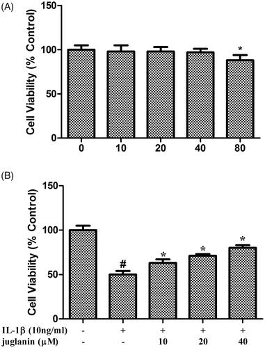
Figure 2. Juglanin decreased iNOS and COX-2 expression in IL-1β-induced human OA chondrocytes. The cells were pretreated with various concentrations of juglanin (10, 20 and 40 μM) for 2 h before subsequent IL-1β stimulation for 24 h. NO concentration in the culture medium was determined by the Griess reaction (A). PGE2 concentration was determined by an Enzyme-linked immunosorbent assay (ELISA) kit (B). Data are expressed as mean ± SEM. All experiments were repeated three times. #p < .05 compared to control group. *p < .05 compared to IL-1β treatment group.
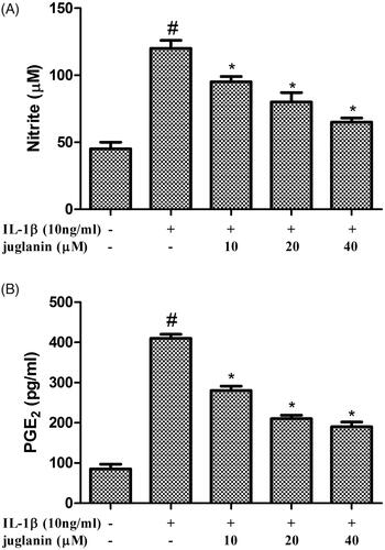
Figure 3. Juglanin decreased iNOS and COX-2 expression in IL-1β-induced human OA chondrocytes. The cells were pretreated with various concentrations of juglanin (10, 20 and 40 μM) for 2 h before subsequent IL-1β stimulation for 24 h. The expression of these proteins and mRNA were assessed by Western blot and qRT-PCR. Data are expressed as mean ± SEM. All experiments were repeated three times. #p < .05 compared to control group. *p < .05 compared to IL-1β treatment group.
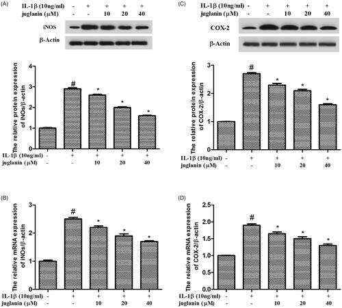
Figure 4. Juglanin inhibited the production of TNF-α and IL-6 in IL-1β-stimulated human OA chondrocytes. The cells were pretreated with various concentrations of juglanin (10, 20 and 40 μM) for 2 h before subsequent IL-1β stimulation for 24 h. TNF-α and IL-6 concentrations were determined by ELISA kits. Data are expressed as mean ± SEM. All experiments were repeated three times. #p < .05 compared to control group. *p < .05 compared to IL-1β treatment group.
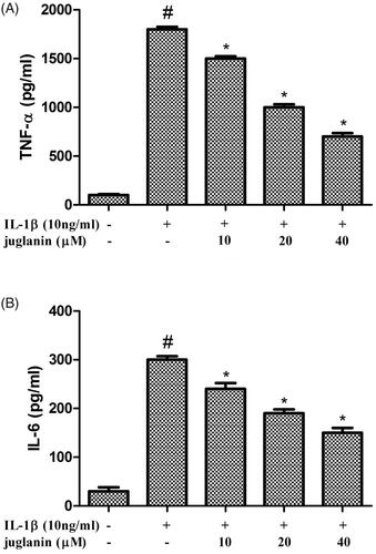
Figure 5. Juglanin decreased production of MMP-1, MMP-3 and MMP-13 in IL-1β-stimulated chondrocytes. The cells were pretreated with various concentrations of juglanin (10, 20 and 40 μM) for 2 h before subsequent IL-1β stimulation for 24 h. MMP-1, MMP-3 and MMP-13 concentrations were determined by ELISA kits. Data are expressed as mean ± SEM. All experiments were repeated three times. #p < .05 compared to control group. *p < .05 compared to IL-1β treatment group.
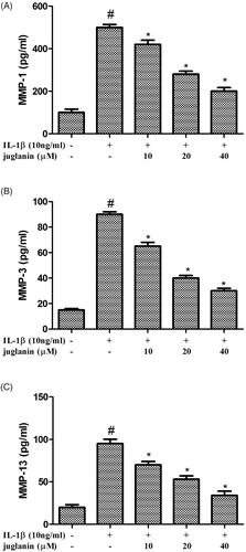
Figure 6. Juglanin suppressed the expression of ADAMTS-4 and ADAMTS-5 in IL-1β-stimulated chondrocytes. The cells were pretreated with various concentrations of juglanin (10, 20 and 40 μM) for 2 h before subsequent IL-1β stimulation for 24 h. The expression of these proteins and mRNA were assessed by Western blot and qRT-PCR. Data are expressed as mean ± SEM. All experiments were repeated three times. #p < .05 compared to control group. *p < .05 compared to IL-1β treatment group.
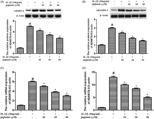
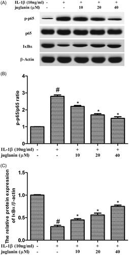
Figure 7. Juglanin prevented IL-1β-induced NF-κB activation in chondrocytes. The cells were pretreated with various concentrations of juglanin (10, 20 and 40 μM) for 2 h before subsequent IL-1β stimulation for 24 h. The expression of these proteins was assessed by Western blot. Data are expressed as mean ± SEM. All experiments were repeated three times. #p < .05 compared to control group. *p < .05 compared to IL-1β treatment group.