
Figures & data
Figure 1. LINC01354 was upregulated in lung cancer. (A) qRT-PCR was used to determine analysis for LINC01354 expression in 43 paired lung cancer tissues. (B) High LINC01354 expression was correlated with advanced TNM stage of patients. (C) LINC01354 expression in lung cancer cells by qRT-PCR. (D, E) LINC01354 expression was significantly reduced in lung tissues by NCBI and UCSC online database. (F, G) High LINC01354 expression predicted a poor overall survival (F) and disease free survival (G). *p < .05.
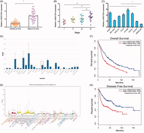
Figure 2. LINC01354 suppression inhibited the proliferation and invasion in lung cancer. (A, B) Relative expression of LINC01354 in lung cancer cells transfected with siRNAs or si-NC. (C-F) CCK8 and colony formation assays were conducted to determine the proliferation in lung cancer. (G, H) Transwell assay was utilized to analyze the invasion in lung cancer. *p < .05.
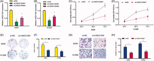
Figure 3. LINC01354 served as a sponge for miR-340-5p. (A, B) LINC01354 location was determined by LncLocator and qRT-PCR. (C-E) Schematic view of miR-340-5p putative binding site in the LINC01354. (F) LINC01354 inhibition reduced miR-340-5p expression in lung cancer cells. (G) LINC01354 expression was negatively correlated with miR-340-5p expression in NSCLC tissues. (H) Dual luciferase reporter assays showed that miR-340-5 bind to LINC01354 and inhibits luciferase activity. (I) RNA pull-down analysis showed a significant amount of LINC01354 and miR-340-5p in the LINC01354 pulled down pellet compared with control group. (J) RIP assay verified the binding sites between miR-340-5p and LINC01354. *p < .05.
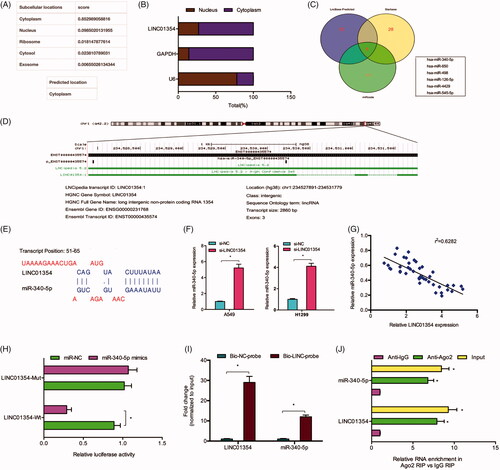
Figure 4. ATF1 acted as a target of miR-340-5p. (A) miR-340-5p expression in 43 paired lung cancer tissues was determined by qRT-PCR. (B, C) Low miR-340-5p expression predicted a poor overall survival rate of patients. (D. E) The predicted complementary binding sites between miR-340-5p and ATF1. (F) miR-340-5p mimics decreased the luciferase activity of ATF1-Wt group by dual-luciferase assay. (G) miR-340-5p mimics reduced the ATF1 protein expression in lung cancer cells. *p < .05.
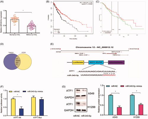
Figure 5. The ATF1 roles in NSCLC. (A) ATF1 expression was upregulated and associated advanced TNM stage in patients with lung cancer. (B) TCGA database showed that ATF1 expression was increased in lung cancer tissues. (C) High ATF1 expression was associated with advanced tumor stage in NSCLC patients. (D-F) High ATF1 expression predicted a poor overall survival rate in NSCLC patients. (G) ATF1 inhibition reduced A549 cells invasion ability in vitro. *p < .05.
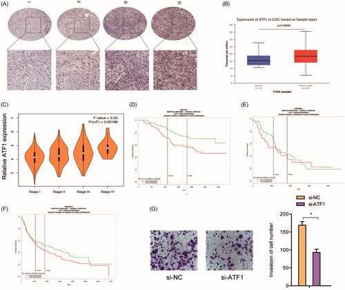
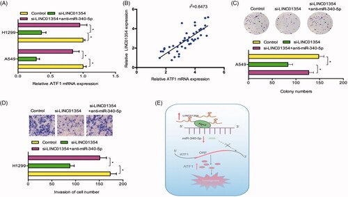
Figure 6. LINC01354/miR-340-5p/ATF1 axis promoted lung cancer progression. (A) miR-340-5p inhibitors rescued the effects of LINC01354 inhibition on ATF1 mRNA expression in lung cancer cells. (B) LINC01354 expression was positively correlated with ATF1 expression in NSCLC tissues. (C, D) LINC01354 suppression on lung cancer cells colony numbers and invasion ability could be rescued by miR-340-5p inhibitors. (E) Schematic diagram of LINC01354/miR-340-5p/ATF1 axis in lung cancer progression *p < .05.
Data availability
The dataset supporting the conclusions of this article is included within the article.