
Figures & data
Table 1. Detailed preparation of the drug delivery system under various treatment conditions.
Figure 1. Photos showing the drug delivery scheme from representative samples 1–5. (a) Nanodiamond–doxorubicin solution after the binding condition is adjusted to pH = 8.0. (b) Pelleting of nanodiamond–doxorubicin complexes after the centrifugation. (c) The resuspended solution after supernatant from part (b) is separated and replaced by an identical volume of a physiological buffer (5 min, 5000 rpm). The final colour is orange.
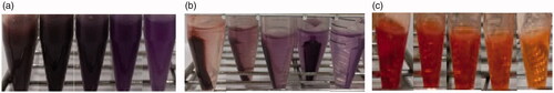
Figure 2. Representative absorbance of 1.0 mg/mL doxorubicin (DOX) before and after adsorption. About 0.2 mg/mL of the added 1.0 mg/mL DOX is remained in the supernatant after facilitating adsorption to nanodiamond–doxorubicin complexes. The absorbance peak of nanodiamond–doxorubicin is found at 495 nm.
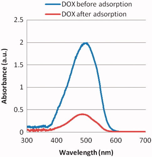
Figure 3. Representative fluorescence intensity of 1.0 mg/mL doxorubicin (DOX) before and after adsorption. The fluorescence peak of nanodiamond–doxorubicin is found at 600 nm.
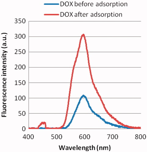
Figure 4. Fluorescence polarization as a function of concentration of nanodiamond–doxorubicin.
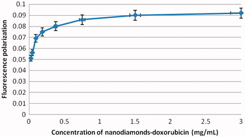
Figure 5. Doxorubicin (DOX) loading capacity and efficacy at various treatment conditions. NDX represents nanodiamond–doxorubicin complexes.
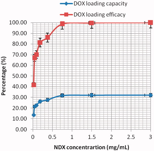
Figure 6. Representative fluorescent microscopy images of MCF-7 cells after 12 h exposure (a) and after 24 h exposure (b) to 3.0 mg/mL nanodiamond–doxorubicin conjugates. Green identifies the fluorescence due to Ethidium Bromide staining. All images are shown with 40× magnification.
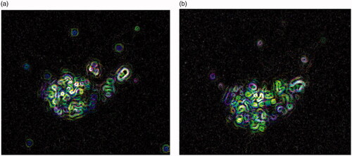
Figure 7. Cytotoxicity of MCF-7 cells after exposure to a range of treatment concentrations. NDX represents nanodiamond–doxorubicin complexes. Dash line denotes IC-50 value.
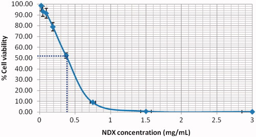
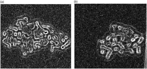
Figure 8. Representative bright-field microscopy images of Vero as the normal cells after 24 h exposure with 5.0 mg/mL nanodiamond alone (a) and 1.0 mg/mL doxorubicin alone (b). The interaction of doxorubicin with Vero was much higher than that of nanodiamond. All images are shown with 40× magnification.
Data availability
All data used to support the findings of this study are included in the article.