Figures & data
Figure 1. H2AT120ph was specifically promoted by Ras-PI3K pathway activation. (A) Empty-pEGFP-N1, pEGFP-RasWT, pEGFP-RasG12V/Y40C and/or H2AT120D or H2AT120A plasmids were transfected into MG63 or U20S cells, respectively. The H2AT120ph expression was measured using western blotting followed by densitometric analysis. (B) Empty-pEGFP-N1, pEGFP-RasWT or pEGFP-RasG12V/Y40C plasmid was transfected into MG63 or U2OS cells, respectively. The phosphate-AKTSer473 expression was measured using western blotting. (C) Empty-pEGFP-N1, pEGFP-RasWT or pEGFP-RasG12V/Y40C plasmid was transfected into MG63 or U2OS cells, respectively. The Ras expression was assessed using western blotting. H2AT120ph: Histone H2A phosphorylated on threonine 120; PI3K: Phosphatidylinositol 3-kinase. *p < .05; **p < .01 (n = 3).
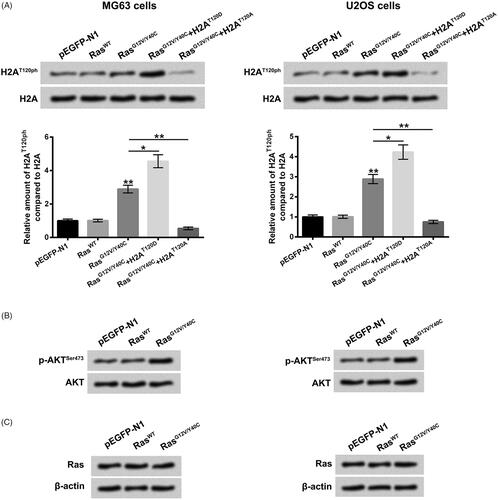
Figure 2. H2AT120ph joined in the oncogenic effects of Ras-PI3K on osteosarcoma. pEGFP-N1, pEGFP-H2A, pEGFP-RasG12V/Y40C and/or pEGFP-H2AT120A were indicated as GFP, H2A, Ras and H2AT120A, respectively. (A) The viabilities of MG63 and U2OS cells were detected using MTT assay after transfection with GFP, H2A, Ras and/or increasing amounts of H2AT120A (0.5, 1 or 2 μM). (B) The numbers of MG63 and U2OS cell colonies were counted after transfection with GFP, H2A, Ras and/or 2 μM H2AT120A. (C) The migration of MG63 and U2OS cells was assessed using two-chamber transwell assay after transfection with GFP, H2A, Ras and/or 2 μM H2AT120A. *p < .05; **p < .01 (n = 3).
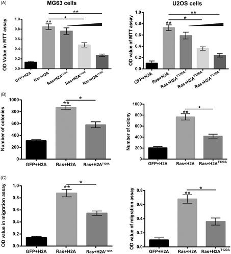
Figure 3. H2AT120D promoted the oncogenic effects of Ras-PI3K on osteosarcoma. pEGFP-N1, pEGFP-H2A, pEGFP-RasG12V/Y40C and/or pEGFP-H2AT120D were indicated as GFP, H2A, Ras and H2AT120D, respectively. (A) The viabilities of MG63 and U2OS cells were detected using MTT assay after transfection with GFP, H2A, Ras and/or 2 μM H2AT120D. (B) The numbers of MG63 and U2OS cell colonies were counted after transfection with GFP, H2A, Ras and/or 2 μM H2AT120D. (C) The migration of MG63 and U2OS cells was assessed using two-chamber transwell assay after transfection with GFP, H2A, Ras and/or 2 μM H2AT120D. *p < .05; **p < .01 (n = 3).
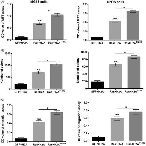
Figure 4. H2AT120ph regulated the transcription of Ras-PI3K-targeted genes in osteosarcoma cells. pEGFP-N1, pEGFP-H2A, pEGFP-RasG12V/Y40C and pEGFP-H2AT120A were indicated as GFP, H2A, Ras and H2AT120A, respectively. (A) The CYR61, IGFBP3, WNT16B, NT5E, GDF15 and CARD16 mRNA expressions in MG63 and U2OS cells were tested using real-time PCR after transfection with GFP, H2A, Ras and/or 2 μM H2AT120A. (B) The input levels of H2AT120ph in promoter regions of CYR61, IGFBP3, WNT16B, NT5E, GDF15 and CARD16 in MG63 cells were evaluated using chromatin immunoprecipitation (ChIP) after transfection with GFP, H2A and/or Ras. *p < .05; **p < .01 (n = 3).
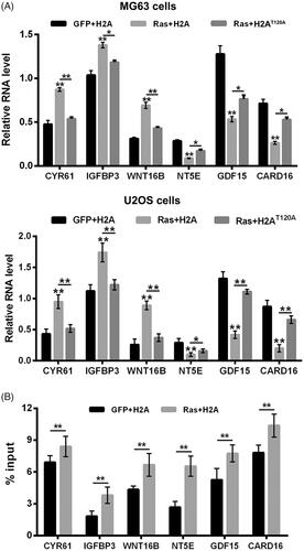
Figure 5. Suppression of VRK1 alleviated Ras-PI3K-induced up-regulation of H2AT120ph and suppressed osteosarcoma progression. (A) After siNC or si-VRK1 transfection, the VRK1 protein level in MG63 cells was detected using western blotting. (B) After pEGFP-N1, pEGFP-RasG12V/Y40C, si-VRK1 and/or siNC transfection, the H2AT120ph expression in MG63 cells was measured using western blotting. pEGFP-N1, and pEGFP-RasG12V/Y40C were indicated as GFP and Ras, respectively. After GFP, Ras, siNC and/or si-VRK1 transfection, (C) the viability of MG63 cells was detected using MTT assay; (D) the migration of MG63 cells was measured using two-chamber transwell assay; (E) the cell cycle distribution of MG63 cells was assessed using flow cytometric analysis. VRK1: Vaccinia-related kinase 1. *p < .05; **p < .01 (n = 3).
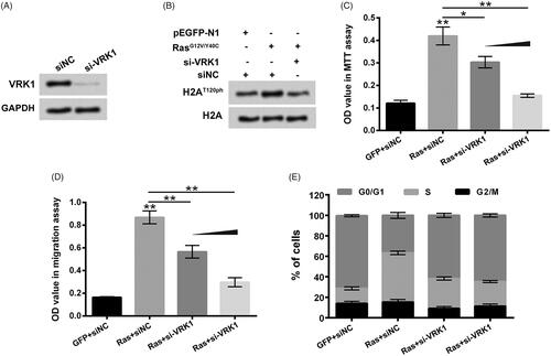
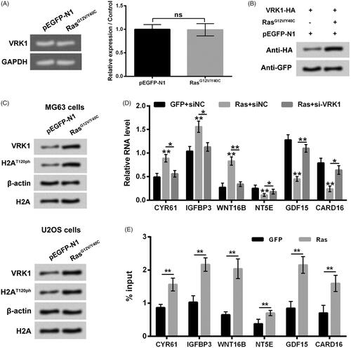
Figure 6. Ras-PI3K pathway specific activation-induced up-regulation of H2AT120ph was achieved by up-regulation of VRK1. (A, B) After pEGFP-N1 or pEGFP-RasG12V/Y40C transfection, the VRK1 mRNA and protein levels were detected using real-time PCR and western blotting, respectively. (C) After pEGFP-N1 or pEGFP-RasG12V/Y40C transfection, the VRK1 and H2AT120ph expression in MG63 and U2OS cells were measured using western blotting. (D) The CYR61, IGFBP3, WNT16B, NT5E, GDF15 and CARD16 mRNA expressions in MG63 cells were tested using real-time PCR after transfection with pEGFP-N1, pEGFP-RasG12V/Y40C and/or siNC or si-VRK1. (E) The input levels of VRK1 in promoter regions of CYR61, IGFBP3, WNT16B, NT5E, GDF15 and CARD16 in MG63 cells were evaluated using chromatin immunoprecipitation (ChIP) after transfection with pEGFP-N1 or pEGFP-RasG12V/Y40C. VRK1: Vaccinia-related kinase 1. **p < .01 (n = 3).