Figures & data
Figure 1. Expression profiles of LINC01518 and miR-1-3p in ESCC cells. qRT-PCR analysis of LINC01518 (A) and miR-1-3p (B) in ESCC cell lines (Ec9706, Eca109, TE-1 and KYSE140) and normal human oesophageal epithelial cell line (Het-1A). (C) The expression of LINC01518 in Ec9706 and TE-1 cells after transfection with si-LINC01518 or si-ctrl for 48 h was examined by qRT-PCR. (D) The expression of miR-1-3p in Ec9706 and TE-1 cells was determined by qRT-PCR 48 h after transfection with miR-1-3p or miR-ctrl. *p < .05.
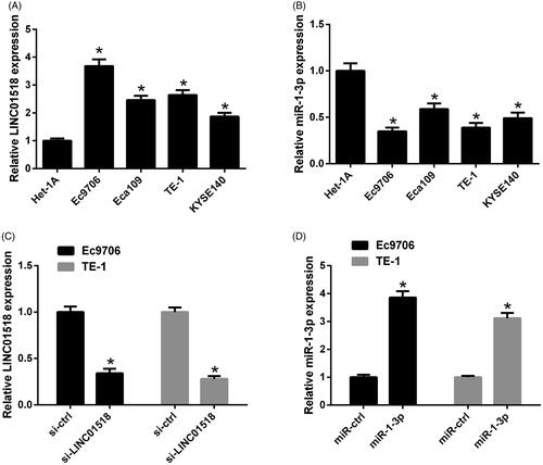
Figure 2. Effect of LINC01518 knockdown on cell proliferation and apoptosis in ESCC cells. (A and B) Ec9706 and TE-1 cells were transfected with si-LINC01518 or si-ctrl for 0, 24, 48, and 72 h, followed by assessment of cell proliferation by CCK-8 assay. (C and D) Ec9706 and TE-1 cells were transfected with si-LINC01518 or si-ctrl for 48 h, followed by the assessment of apoptosis by flow cytometry analysis. *p < .05.
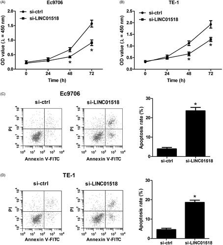
Figure 3. The interaction between LINC01518 and miR-1-3p in ESCC cells. (A) The wild-type and mutated sequences of LINC01518 containing binding sequences complementary to miR-1-3p seed regions. (B) Luciferase activity was measured by luciferase activity assay in Ec9706 and TE-1 cells 48 h after cotransfection with LINC01518-WT or LINC01518-MUT and miR-1-3p or miR-ctrl. (C) miR-1-3p expression was estimated in Ec9706 and TE-1 cells 48 h after transfection with si-LINC01518 or si-ctrl. *p < .05.
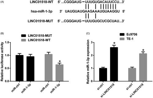
Figure 4. Effects of LINC01518 silencing or combined with miR-1-3p downregulation on cell proliferation and apoptosis in ESCC cells. (A and B) Ec9706 and TE-1 cells were cotransfected with si-LINC01518 or si-ctrl and anti-miR-1-3p or anti-miR-ctrl for 0, 24, 48, and 72 h, followed by the assessment of cell proliferation by CCK-8 assay. (C and D) Ec9706 and TE-1 cells were cotransfected with si-LINC01518 or si-ctrl and anti-miR-1-3p or anti-miR-ctrl for 48 h, followed by the assessment of apoptosis by flow cytometry analysis. *p < .05.
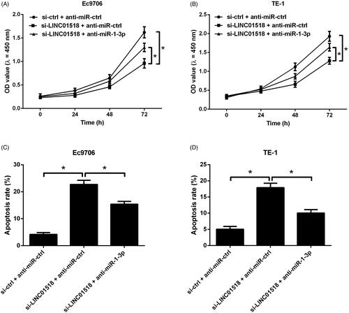
Figure 5. The interaction between PIK3CA and miR-1-3p in ESCC cells. (A) Schematic of the wild-type or mutated PIK3CA 3′UTR containing miR-1-3p binding sites. (B) Luciferase activity was measured by luciferase reporter assay 48 h after Ec9706 cells were cotransfected with PIK3CA-WT or PIK3CA-MUT and miR-1-3p or miR-ctrl. (C) The protein levels of PIK3CA, p-Akt and Akt were detected by Western blot in Ec9706 and TE-1 cells 48 h after transfection with miR-1-3p or miR-ctrl. *p < .05.
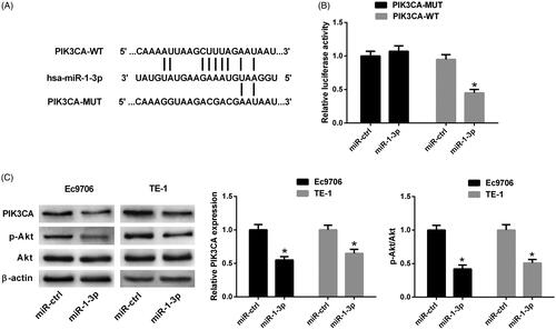
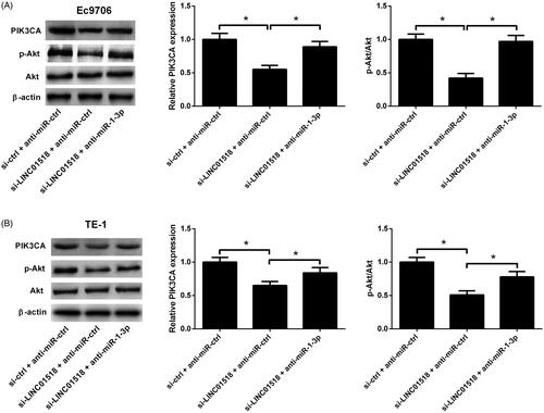
Figure 6. Effects of LINC01518 knockdown or along with anti-miR-1-3p on the PIK3CA/Akt pathway in ESCC cells. (A and B) Ec9706 and TE-1 cells were cotransfected with si-LINC01518 or si-ctrl and anti-miR-1-3p or anti-miR-ctrl for 48 h, followed by Western blot analysis of PIK3CA, p-Akt and Akt protein levels. *p < .05.