
Figures & data
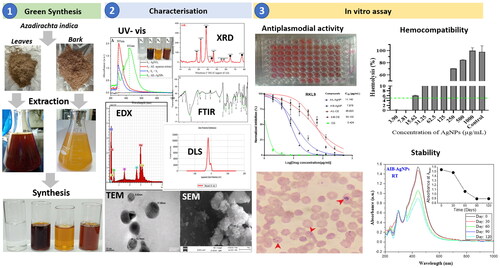
Figure 1. Optimization of biosynthesis AgNPs using A. indica leaves and bark aqueous extract by monitoring UV–Visible absorption spectra: Effects of incubation time (A: leaves; B: bark); AgNO3 concentration (C: leaves; D: bark); Extract concentration (E: leaves; F: bark) and Temperature (G: leaves; H: bark).
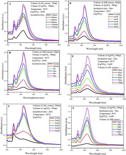
Figure 1. Continued.
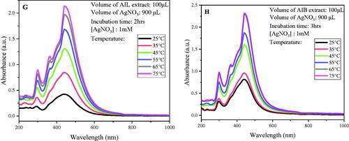
Figure 2. UV–vis absorption spectra of silver nitrate (S1), aqueous extracts (S2), Silver nitrate and aqueous extract mixture at T0 (S3) and biosynthesized silver nanoparticles (S4) using A. indica leaves (A) and Bark (B).
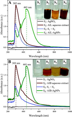
Figure 3. X-ray diffraction pattern of silver nanoparticles from A. indica leaves (AIL) and bark (AIB). ● represents silver nanocrystallites and ![]()
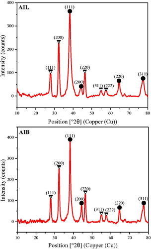
Figure 4. Energy-dispersive X-ray profile of A. indica leaves (A) and bark (B) silver nanoparticles (AgNPs) showing their elemental composition. Scanning electron microscopy images of A. indica leaves (C) and bark (D) AgNPs.
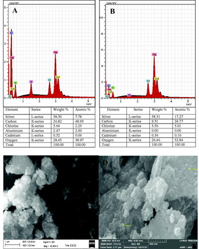
Figure 5. Transmission electron microscopy images of biosynthesized silver nanoparticles (AgNPs) from A. indica leaves (A) and bark (B). Selected area electron diffraction pattern showing multiple diffraction rings indicating polycrystalline nature of AgNPs leaves (C) and bark (D). Particles size distribution of the AgNPs leaves (E) and bark (F).
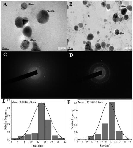
Figure 6. FTIR spectra of AgNPs synthesized from A. indica leaves (AIL) and Bark (AIB).
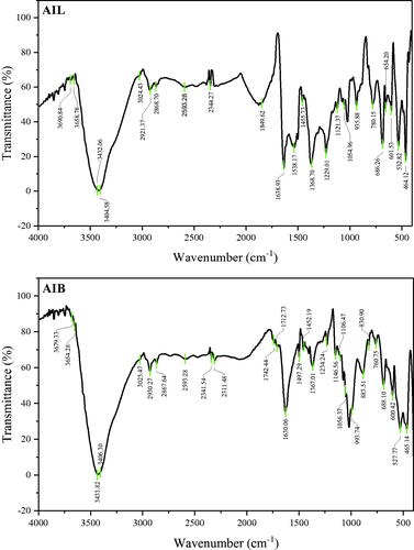
Table 1. FTIR profile of silver nanoparticles synthesized from A. indica leaves and bark.
Table 2. Comparison of some characteristics with previous studies that reported antiplasmodial activity of silver nanoparticles synthesised from A. indica.
Figure 7. Anti-plasmodial activity of Chloroquine (CQ), A. indica leaves and Bark aqueous extract and their corresponding silver nanoparticle against CQ-sensitive strain (3D7) and CQ-Resistant strain (RKL9). AIL-AgNP: A. indica leaves silver nanoparticles; AIB-AgNP: A. indica bark silver nanoparticles; AIL-CE: A. indica leaves crude aqueous extract; AIB-CE: A. indica bark crude aqueous extract.
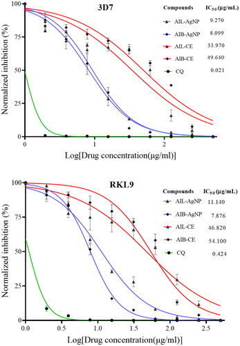
Figure 8. (A) Aspect of culture after synchronization showing healthy parasite at ring stage. Same synchronized culture was used for all tested compounds. (B) Aspect of culture after 24 and 48 h of incubation with different drugs. CQ: Chloroquine; AIL-AgNPs: A. indica leaves silver nanoparticles, AIB-AgNPs: A. indica bark silver nanoparticles.
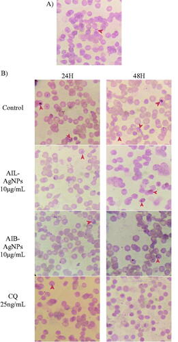
Table 3. Antiplasmodial activity of aqueous extract and A. indica AgNPs after 120 days of storage.
Figure 9. Haemolysis potential of human red blood cells of synthesized A. indica silver nanoparticles leaves (A–D) and bark (E–H). Triton X (10%) was used as control. Results are plotted as normalized mean ± standard deviation. The green dash line represents a 5% cut off line according to the ASTM criteria (E2524-08 card). Fluorescence quenching effects of aqueous extracts and silver nanoparticles using 3D7 (I) and RKL9 (J) strains. AIL-AgNPs: A. indica leaves silver nanoparticles; AIB-AgNPs: A. indica bark silver nanoparticles; AIL-CE: A. indica leaves crude extract; AIB-CE: A. indica bark crude extract.
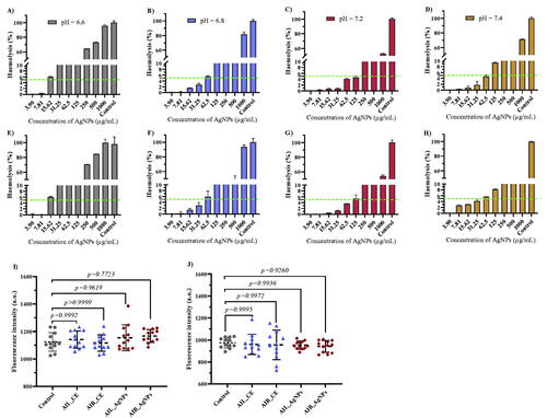
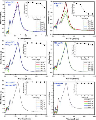
Figure 10. Surface plasma resonance stability of silver nanoparticles in different storage condition. AIL-AgNPs: A. indica leaves silver nanoparticles; AIB-AgNPs: A. indica bark silver nanoparticles; RT: room temperature.
Supplemental Material
Download MS Word (135.3 KB)Data availability statement
The authors confirm that the data supporting the findings of this study are available within the article and in supplementary files.