
Figures & data
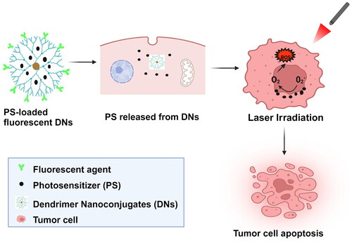
Figure 1. Schematic representation of active and passive targeted drug delivery in anticancer therapy – reprinted from reference [Citation63], copyright 2023, Springer Nature.
![Figure 1. Schematic representation of active and passive targeted drug delivery in anticancer therapy – reprinted from reference [Citation63], copyright 2023, Springer Nature.](/cms/asset/0126e451-74ac-43a7-8fde-095188812a8d/ianb_a_2368033_f0001_c.jpg)
Table 1. Types of mechanisms in which dendrimers penetrate tumour cells.
Figure 2. In vitro fluorescence or confocal imaging of dendrimer-based nanoparticles in cancer cells. (i) Confocal images of SH-SY5Y cells incubated for 20 min with curcumin/resveratrol/dendrimer NPs – reprinted from reference [Citation89], copyright 2022, ACS publications; (ii) sugar-conjugated dendrimer localizations in glioblastoma – reprinted from reference [Citation90], copyright 2021, Elsevier; and (iii) PEGylated bis-indolyl G2 and G3 polyurethane dendrimer treated cells (A) MDA-MB-231 cells and (B) A549 cells – reprinted from reference [Citation91], copyright 2021, Elsevier.
![Figure 2. In vitro fluorescence or confocal imaging of dendrimer-based nanoparticles in cancer cells. (i) Confocal images of SH-SY5Y cells incubated for 20 min with curcumin/resveratrol/dendrimer NPs – reprinted from reference [Citation89], copyright 2022, ACS publications; (ii) sugar-conjugated dendrimer localizations in glioblastoma – reprinted from reference [Citation90], copyright 2021, Elsevier; and (iii) PEGylated bis-indolyl G2 and G3 polyurethane dendrimer treated cells (A) MDA-MB-231 cells and (B) A549 cells – reprinted from reference [Citation91], copyright 2021, Elsevier.](/cms/asset/eb57cb72-8264-459e-8706-15bab633730e/ianb_a_2368033_f0002_c.jpg)
Figure 3. In vivo MRI images of dendrimer-based NPs in cancer model (i) (a) the coronal MRI images shows the location of U-251 glioma tumour, (b) distribution of G3-curcumin in organs and (c) fluorescence images of kidney (K), spleen (Sp), liver, heart (H), lung (L) and brain and (B) in rat model – reprinted from reference [Citation92], copyright 2016, PubMed Central; and (ii) G5-Gd trastuzumab and G5-Gd NPs injected tumour bearing mice model – reprinted from reference [Citation93], copyright 2020, Springer Nature).
![Figure 3. In vivo MRI images of dendrimer-based NPs in cancer model (i) (a) the coronal MRI images shows the location of U-251 glioma tumour, (b) distribution of G3-curcumin in organs and (c) fluorescence images of kidney (K), spleen (Sp), liver, heart (H), lung (L) and brain and (B) in rat model – reprinted from reference [Citation92], copyright 2016, PubMed Central; and (ii) G5-Gd trastuzumab and G5-Gd NPs injected tumour bearing mice model – reprinted from reference [Citation93], copyright 2020, Springer Nature).](/cms/asset/94ccdda8-03a9-442c-9760-4743a73808b8/ianb_a_2368033_f0003_c.jpg)
Table 2. Fundamentals of dendrimer-based nanoconjugates for tumour elimination.
Figure 4. [a] Biodegradation and [b] Clearance properties of a dendrimer.
![Figure 4. [a] Biodegradation and [b] Clearance properties of a dendrimer.](/cms/asset/f72c6242-30d6-4f39-be45-8088fddf786a/ianb_a_2368033_f0004_c.jpg)
Data availability statement
Data are openly available in a public repository that issues datasets with DOIs.