3,453
Views
4
CrossRef citations to date
0
Altmetric
Ebola

High dose of vesicular stomatitis virus-vectored Ebola virus vaccine causes vesicular disease in swine without horizontal transmission

, , , , , ORCID Icon, , , , , , , ORCID Icon, ORCID Icon, , , , & ORCID Icon show all
Pages 651-663 | Received 01 Dec 2020, Accepted 09 Mar 2021, Published online: 02 Apr 2021

Figures & data

Figure 1. Clinical analysis of wtVSV-inoculated pigs. (A) Average daily temperatures of wtVSV-inoculated pigs compared to uninoculated pigs. (B) Average lesion scores of wtVSV-inoculated pigs compared to uninoculated environmental controls on each day of the study. (C) Summary of RT-qPCR analysis performed to detect viral RNA in clinical samples collected throughout the study, indicating the presence (+) or absence (−) of viral RNA. P-LN – parotid lymph node; RP-LN – retropharyngeal lymph node; SC-LN – superficial cervical lymph node; M-LN – mandibular lymph node; NP – nasal planum; NA – Not Applicable because not present or collected; DPI - days post inoculation in which positive samples were detected. (D) Representative pictures showing vesicular lesions in pigs. (E) Immunohistochemistry analysis performed on nasal planum lesion tissue collected on 2 DPI necropsy from wtVSV-infected pig #3 showing positive (red) immunostaining using anti-VSV-G rabbit polyclonal antibody localized to the stratum spinosum and granulosum of the epidermis.
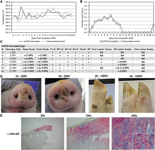
Figure 1. Clinical analysis of wtVSV-inoculated pigs. (A) Average daily temperatures of wtVSV-inoculated pigs compared to uninoculated pigs. (B) Average lesion scores of wtVSV-inoculated pigs compared to uninoculated environmental controls on each day of the study. (C) Summary of RT-qPCR analysis performed to detect viral RNA in clinical samples collected throughout the study, indicating the presence (+) or absence (−) of viral RNA. P-LN – parotid lymph node; RP-LN – retropharyngeal lymph node; SC-LN – superficial cervical lymph node; M-LN – mandibular lymph node; NP – nasal planum; NA – Not Applicable because not present or collected; DPI - days post inoculation in which positive samples were detected. (D) Representative pictures showing vesicular lesions in pigs. (E) Immunohistochemistry analysis performed on nasal planum lesion tissue collected on 2 DPI necropsy from wtVSV-infected pig #3 showing positive (red) immunostaining using anti-VSV-G rabbit polyclonal antibody localized to the stratum spinosum and granulosum of the epidermis.

Figure 2. Clinical analysis of VSV-EBOV-inoculated pigs. (A) Average daily temperatures of VSV-EBOV-inoculated pigs compared to uninoculated controls. (B) Average lesion scores of VSV-EBOV-inoculated pigs compared to uninoculated controls on each day of the study. (C) Summary of RT-qPCR analysis performed to detect viral RNA in clinical samples collected throughout the study, indicating the presence (+) or absence (−) of viral RNA. P-LN – parotid lymph node; RP-LN – retropharyngeal lymph node; SC-LN – superficial cervical lymph node; M-LN – mandibular lymph node; NP – nasal planum; NA – Not Applicable because not present or collected; DPI - days post inoculation in which positive samples were detected. (D) Representative pictures showing vesicular lesions in VSV-EBOV-inoculated pigs. (E) Immunohistochemistry analysis performed on foot lesion tissue (hoof and skin) collected on 10 DPI necropsy from VSV-EBOV-inoculated pig #15 showing positive (red) immunostaining using anti-EBOV-GP rabbit polyclonal antibody localized to the stratum spinosum and granulosum of the epidermis.
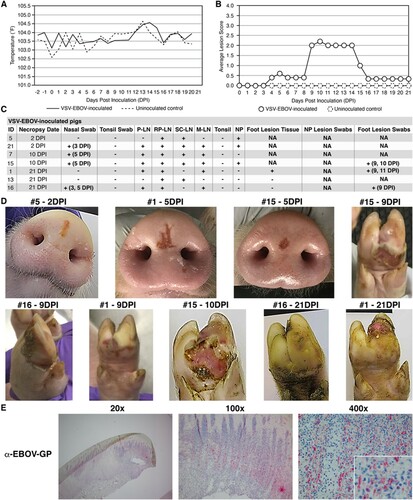
Figure 2. Clinical analysis of VSV-EBOV-inoculated pigs. (A) Average daily temperatures of VSV-EBOV-inoculated pigs compared to uninoculated controls. (B) Average lesion scores of VSV-EBOV-inoculated pigs compared to uninoculated controls on each day of the study. (C) Summary of RT-qPCR analysis performed to detect viral RNA in clinical samples collected throughout the study, indicating the presence (+) or absence (−) of viral RNA. P-LN – parotid lymph node; RP-LN – retropharyngeal lymph node; SC-LN – superficial cervical lymph node; M-LN – mandibular lymph node; NP – nasal planum; NA – Not Applicable because not present or collected; DPI - days post inoculation in which positive samples were detected. (D) Representative pictures showing vesicular lesions in VSV-EBOV-inoculated pigs. (E) Immunohistochemistry analysis performed on foot lesion tissue (hoof and skin) collected on 10 DPI necropsy from VSV-EBOV-inoculated pig #15 showing positive (red) immunostaining using anti-EBOV-GP rabbit polyclonal antibody localized to the stratum spinosum and granulosum of the epidermis.

Figure 3. Clinical analysis of wtVSV-contact control pigs. (A) Average daily temperatures of wtVSV- contact control pigs compared to uninoculated controls. (B) Average lesion scores of wtVSV-contact control pigs compared to uninoculated controls on each day of the study. (C) Summary of RT-qPCR analysis performed to detect viral RNA in clinical samples collected throughout the study, indicating the presence (+) or absence (−) of viral RNA. P-LN – parotid lymph node; RP-LN – retropharyngeal lymph node; SC-LN – superficial cervical lymph node; M-LN – mandibular lymph node; NP – nasal planum; NA – Not Applicable because not present or collected; DPI - days post inoculation of principal infected pigs in which positive samples were detected in this group. (D) Representative pictures showing vesicular lesions in wtVSV-contact control pigs.
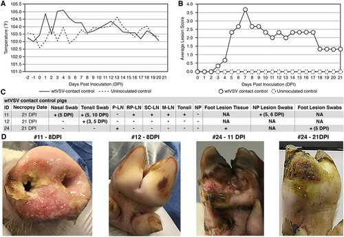
Figure 3. Clinical analysis of wtVSV-contact control pigs. (A) Average daily temperatures of wtVSV- contact control pigs compared to uninoculated controls. (B) Average lesion scores of wtVSV-contact control pigs compared to uninoculated controls on each day of the study. (C) Summary of RT-qPCR analysis performed to detect viral RNA in clinical samples collected throughout the study, indicating the presence (+) or absence (−) of viral RNA. P-LN – parotid lymph node; RP-LN – retropharyngeal lymph node; SC-LN – superficial cervical lymph node; M-LN – mandibular lymph node; NP – nasal planum; NA – Not Applicable because not present or collected; DPI - days post inoculation of principal infected pigs in which positive samples were detected in this group. (D) Representative pictures showing vesicular lesions in wtVSV-contact control pigs.

Figure 4. Clinical analysis of VSV-EBOV-contact control pigs. (A) Average daily temperatures of VSV-EBOV contact control pigs compared to uninoculated controls. (B) Average lesion scores of VSV-EBOV contact control pigs compared to uninoculated controls on each day of the study. * – one pig (#10) showed a focal <2 cm reddening on a hind limb on 14 DPI that was transient and did not show vesicular or ulcerative features typical of VSV infection and was ultimately determined to be a mechanical injury. (C) Summary of RT-qPCR analysis performed to detect viral RNA in clinical samples collected throughout the study, indicating the presence (+) or absence (−) of viral RNA. P-LN – parotid lymph node; RP-LN – retropharyngeal lymph node; SC-LN – superficial cervical lymph node; M-LN – mandibular lymph node; NP – nasal planum; NA – Not Applicable because lesions not present or collected; DPI - days post inoculation of principal infected pigs. * – suspected foot lesion swabs and suspected lesion tissues were collected on any tissue that was discolored even if the cause was likely a mechanical injury or benign discoloration and tested for viral RNA. All such samples were negative for VSV-specific RNA. (D) Representative pictures showing a lack of vesicular lesions in VSV-EBOV-contact control pigs.
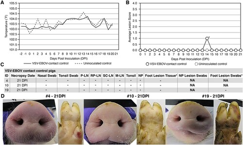
Figure 4. Clinical analysis of VSV-EBOV-contact control pigs. (A) Average daily temperatures of VSV-EBOV contact control pigs compared to uninoculated controls. (B) Average lesion scores of VSV-EBOV contact control pigs compared to uninoculated controls on each day of the study. * – one pig (#10) showed a focal <2 cm reddening on a hind limb on 14 DPI that was transient and did not show vesicular or ulcerative features typical of VSV infection and was ultimately determined to be a mechanical injury. (C) Summary of RT-qPCR analysis performed to detect viral RNA in clinical samples collected throughout the study, indicating the presence (+) or absence (−) of viral RNA. P-LN – parotid lymph node; RP-LN – retropharyngeal lymph node; SC-LN – superficial cervical lymph node; M-LN – mandibular lymph node; NP – nasal planum; NA – Not Applicable because lesions not present or collected; DPI - days post inoculation of principal infected pigs. * – suspected foot lesion swabs and suspected lesion tissues were collected on any tissue that was discolored even if the cause was likely a mechanical injury or benign discoloration and tested for viral RNA. All such samples were negative for VSV-specific RNA. (D) Representative pictures showing a lack of vesicular lesions in VSV-EBOV-contact control pigs.
Supplemental material

Supplemental Material

Download PDF (4.6 MB)