Figures & data
Figure 1. Flow chart of all patients who underwent EGD in the study period and selection of study population.
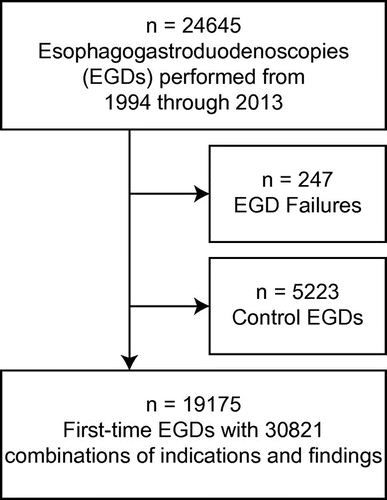
Table 1. Frequency of findings in 30,821 pairs of indications and findings in 19,175 EGDs
Figure 2. Age distribution of findings with limited or no implication (solid lines) and findings with or possibly with implication (dashed lines) in females (a) and males (b).
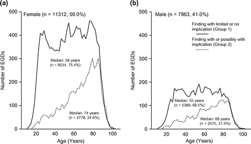
Figure 3. Rate of patients who underwent EGD in different five-year periods (females, solid line; males, dashed line).
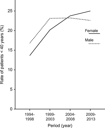
Table 2. Findings in EGDs with the indication of suspicious malignancy (n = 599)
Figure 4. Nomogram predicting finding with limited or no implication upon EGD. Add points for age (1) and points for indication by sex (2) to determine total points (1 + 2). The probability of finding with limited or no implication can be read by matching with the total point scale.
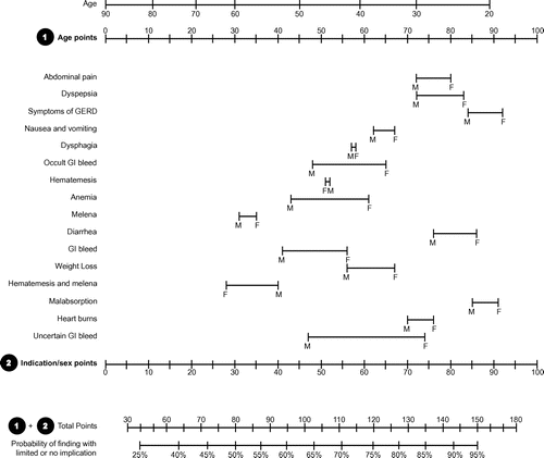
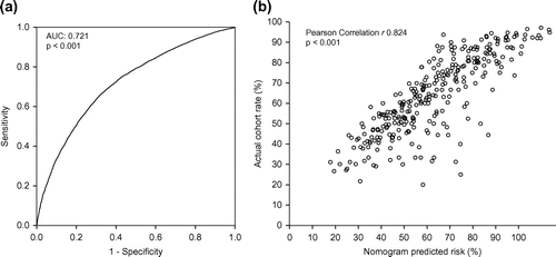
Figure 5. ROC analysis of the nomogram-calculated probability (a) and scatter plot (b) where each circle represents a pair of nomogram-calculated probability of finding with limited or no clinical implication and the actual finding rate within each subgroup (n = 256) of age and sex/indication combinations.