Figures & data
Table 1. Mutationen in der Fc-region von IgG
Figure 1. Analysis of immune complexes from synovial fluid of a patient with rheumatoid arthritis. (A) 10% polyacrylamide gel stained with Commassie Blue. (B) Western blot with homologous biotinylated IgG.
Note: The results were representative of 5 preparations.
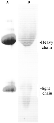
Figure 2. Kinetics of immune complex formation. (A) IgG was isolated from synovial immune complexes of a patient with rheumatoid arthritis by Protein A-Sepharose affinity chromatography. Polyethylene glycol was added to a final concentration of 5%, the acid eluate was neutralized and the turbidity at 630 nm was recorded at room temperature. (B) Size of immune complexes. Particle diameters were determined by nephelometry after neutralization of the Protein A-Sepharose eluate. The weight percents are recorded for the indicated diameters.
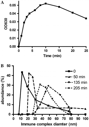
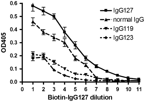
Figure 3. Specificity of rheumatoid antibodies. Immune complexes were isolated from synovial fluid of patients with rheumatoid arthritis.
Notes: One portion of the immune complexes from patient #127 was biotinylated and used as paratope. Another portion from the same patient #127 immune complexes from two other patients #119 and #123 and normal IgG were used as epitope and adsorbed at pH 2.5 to microtiter plate. Biotinylated IgG127 was serially diluted 1:2 and the binding affinity was determined in an ELISA assay. The error bars indicate the range of two determinations.