
Figures & data
Figure 1. Metabolic power (MP; W) at an external power output of 50% POpeak across tests 1, 2 and 3 among adult (n = 9) and adolescent cyclists (n = 9).
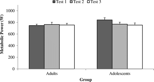
Figure 2. External power output (PO; W) at 50% POpeak across tests 1, 2 and 3 among adult (n = 9) and adolescent cyclists (n = 9).
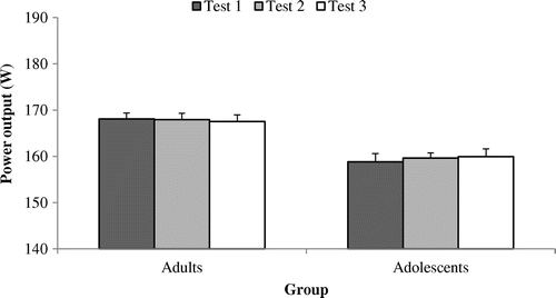
Figure 3. Gross efficiency (GE%) at a power output of 50% POpeak across tests 1, 2 and 3 among adult (n = 9) and adolescent cyclists (n = 9).
Note: † = Significantly different to test l for that group.
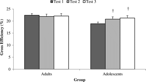
Figure 4. Cadence (rev·min−1) at an external power output of 50% POpeak across tests 1, 2 and 3 among adult (n = 9) and adolescent cyclists (n = 9).
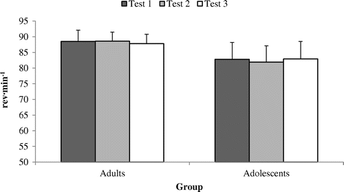
Figure 5. Respiratory exchange ratio (RER) at an external power output of 50% POpeak across tests 1, 2 and 3 among adult (n = 9) and adolescent cyclists (n = 9).
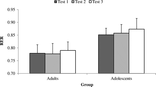
Figure 6. EMG root mean square of the vastus lateralis (VL-EMG-rms [%]) at a power output of 50% POpeak across tests 1, 2 and 3 among adult (n = 9) and adolescent cyclists (n = 9).
Note: † = Significantly different to test l for that group.
![Figure 6. EMG root mean square of the vastus lateralis (VL-EMG-rms [%]) at a power output of 50% POpeak across tests 1, 2 and 3 among adult (n = 9) and adolescent cyclists (n = 9).Note: † = Significantly different to test l for that group.](/cms/asset/abf91614-fd06-4967-93cf-146246edbacb/oamd_a_1237606_f0006_b.gif)
Figure 7. EMG root mean square of the biceps femoris (BF-EMG-rms [%]) at a power output of 50% POpeak across tests 1, 2 and 3 among adult (n = 9) and adolescent cyclists (n = 9).
Note: † = Significantly different to test l for that group.
![Figure 7. EMG root mean square of the biceps femoris (BF-EMG-rms [%]) at a power output of 50% POpeak across tests 1, 2 and 3 among adult (n = 9) and adolescent cyclists (n = 9).Note: † = Significantly different to test l for that group.](/cms/asset/872386a7-2816-4e8e-941b-475b069cd4c8/oamd_a_1237606_f0007_b.gif)
Table 1. Reliability of GE, VL-EMG-rms (%), BF-EMG-rms (%) among adult (n = 9) and adolescent cyclists (n = 9)
Table 2. The relationship (r-values) between the inter-test differences of GE and inter-test differences EMG-rms of the vastus lateralis and biceps femoris among adults (n = 9) and adolescents (n = 9)