Figures & data
Figure 1. Plot of PAI-1 marker concentration in BPH and PCa biopsy tissues against the mean age of patients: (A) initial data (Serafin et al., Citation2016) and (B) validation data.
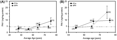
Figure 2. (A) Comparison of malignancy indices in biopsy tissue from BPH (n = 103) and PCa (n = 114) patients. p < 0.05 indicates a statistically significant difference. (B) Plot of malignancy index against the mean age of patients grouped in 10-year intervals. “Other” refers to data from individuals presenting with non-BPH and non-PCa pathologies.
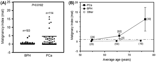
Figure 3. (A) Comparison of malignancy indices in TURP tissue from BPH (n = 70) and PCa (n = 30) patients. p < 0.05 indicates a statistically significant difference. (B) Plot of malignancy index against the mean age of patients grouped in 10-year intervals.
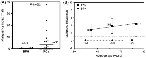
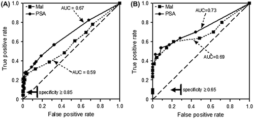
Figure 4. Comparison of ROC curves for malignancy index (Mal) and PSA level. (A) Mal obtained from needle biopsies of 217 patients. (B) Mal obtained from TURP tissues of 100 patients. PSA was routinely measured.