1,548
Views
4
CrossRef citations to date
0
Altmetric
Research Article

Assessment of uterine fibroid infarction after embolization with tris-acryl gelatin microspheres

, , & | (Reviewing Editor)
Article: 1360543 | Received 29 Jun 2017, Accepted 24 Jul 2017, Published online: 03 Aug 2017

Figures & data

Figure 1. (A) Presents contrast-enhanced MRI of the fibroid prior to UFE. (B) Presents the same fibroid after UFE with complete fibroid infarction (100%) without residual contrast enhancement. Category 1.
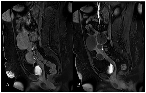
Figure 1. (A) Presents contrast-enhanced MRI of the fibroid prior to UFE. (B) Presents the same fibroid after UFE with complete fibroid infarction (100%) without residual contrast enhancement. Category 1.

Figure 2. (A) Presents contrast-enhanced MRI of the fibroid prior to UFE. (B) Presents the same fibroid after UFE with almost complete fibroid infarction (90–99%) and residual contrast enhancement below 10%. Note small irregularity on the base of embolized fibroid. Category 2.
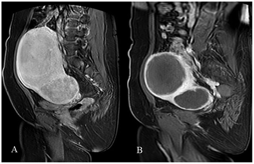
Figure 2. (A) Presents contrast-enhanced MRI of the fibroid prior to UFE. (B) Presents the same fibroid after UFE with almost complete fibroid infarction (90–99%) and residual contrast enhancement below 10%. Note small irregularity on the base of embolized fibroid. Category 2.

Figure 3. (A) Presents contrast-enhanced MRI of the fibroid prior to UFE. (B) present the same fibroid after UFE with incomplete fibroid infarction (<90%) and with residual contrast enhancement over 10%. The central part of the fibroid is with residual contrast enhancement. Category 3.
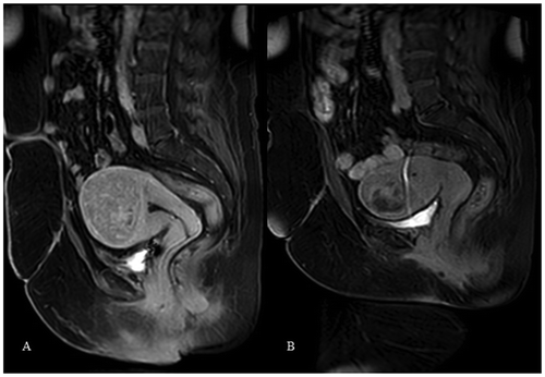
Figure 3. (A) Presents contrast-enhanced MRI of the fibroid prior to UFE. (B) present the same fibroid after UFE with incomplete fibroid infarction (<90%) and with residual contrast enhancement over 10%. The central part of the fibroid is with residual contrast enhancement. Category 3.

Table 1. Patients symptoms and MRI characteristics

Figure 4. (A) Presents fibroid prior to UFE. The left uterine artery was not embolized due to small calibre. (B) Presents almost complete fibroid vascularization from the right uterine artery. (C) Presents control contrast enhancing MRI with complete fibroid infarction despite the left uterine artery was not embolized.
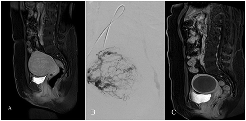
Figure 4. (A) Presents fibroid prior to UFE. The left uterine artery was not embolized due to small calibre. (B) Presents almost complete fibroid vascularization from the right uterine artery. (C) Presents control contrast enhancing MRI with complete fibroid infarction despite the left uterine artery was not embolized.
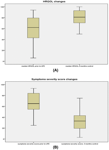
Figure 5. The median changes in the HRQOL and symptoms severity score. (A) Significant increasing in HRQOL after UFE (p < 0.001). (B) Significant decreasing of symptoms severity score (p < 0.001).

Figure 5. The median changes in the HRQOL and symptoms severity score. (A) Significant increasing in HRQOL after UFE (p < 0.001). (B) Significant decreasing of symptoms severity score (p < 0.001).