Figures & data
Figure 1. Ionic interactions of nanoparticle components. (a) A nanoparticle developed previously consisting of RMP, AG-PLL, and PLA with terminal carboxyl groups (PLA-C). (b) A nanoparticle developed in this study consisting of RMP, AG-PLG, and PLA with terminal amino groups (PLA-N1 or PLA-N3). The components in dotted line squares were cationic, and those in solid line squares were anionic. Allows mean ionic interactions. (c) Chemical structures of PLA, RMP, AG-PLL and AG-PLG.
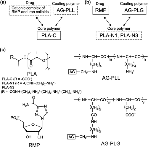
Table 1. Molecular weights of polymers
Table 2. Nanoparticle characteristics
Figure 2. Release behavior of RMP from PLG/N3-NP.
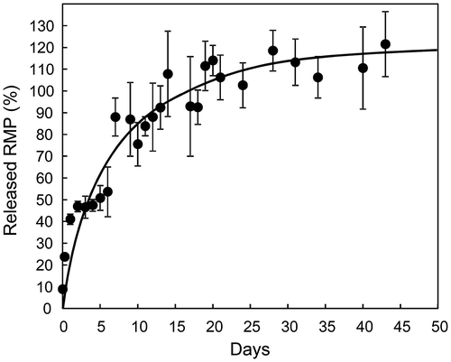
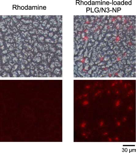
Figure 3. Uptake of rhodamine-loaded nanoparticles in HepG2 cells.
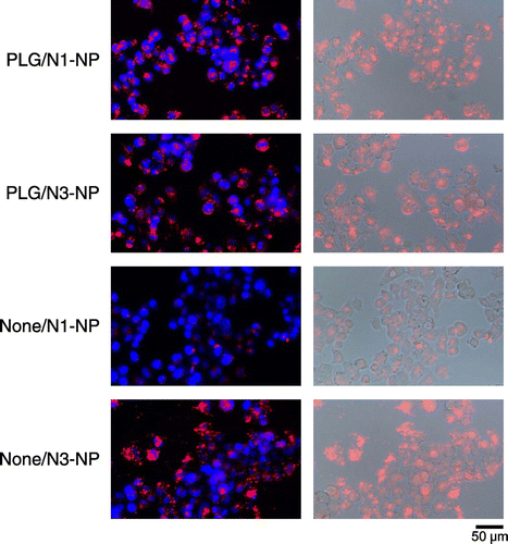
Figure 4. Cytotoxicity of conjugates.
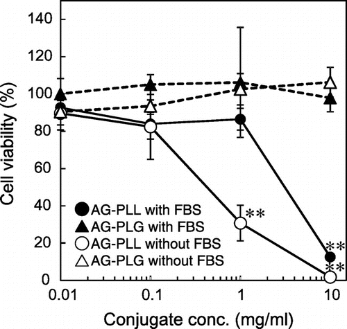
Figure 5. Cytotoxicity of nanoparticles. After incubation of HepG2 cells with various nanoparticles in Opti-MEM for 24 h, (a) cell viability was determined, or (b) the cells were observed using a microscope. Each data point represents the mean ± SD of measurements from three individual wells.
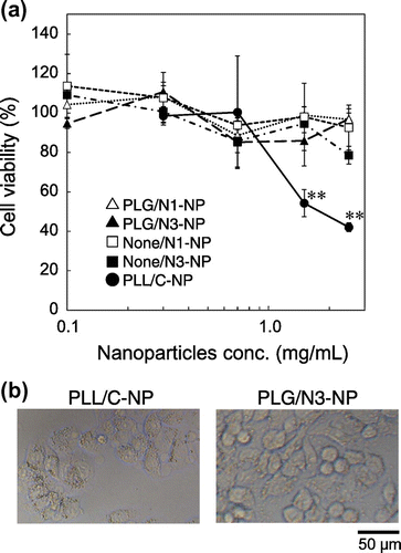
Figure 6. Frozen liver sections of mice.