Figures & data
Table 1. Examples of scientific literature that explore the regeneration of peripheral nerves submitted to axonotmesis with different therapeutic approaches
Figure 1. Schematic representation of the peripheral nerve anatomy and structural overview of the PNS.
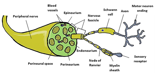
Figure 2. Schematic representation of the different injury grading systems for PNI.
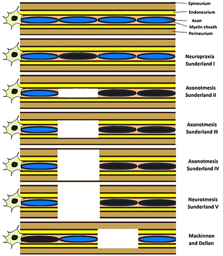
Figure 3. Steps for the induction of an axonotmesis lesion in rat’s sciatic nerve: (a) Isolation of the sciatic nerve from neighboring tissues; (b) induction of injury (material used, time and force of application depends on the selected protocol); (c) anatomical flattening of the sciatic nerve after injury.
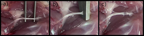
Figure 4. Representative light micrographs of toluidine blue-stained semi-thin cross sections of control and crushed median nerves at different time points in the rat animal model. Bar: 20 μm.
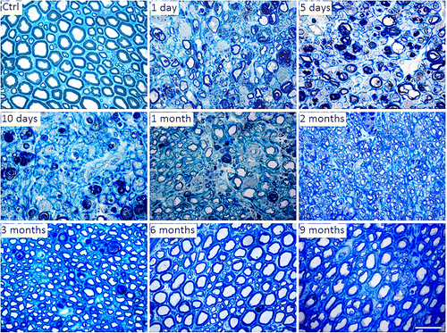
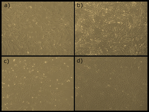
Figure 5. Different types of MSCs in culture, (a) Human Dental Pulp MSCs (P2); (b) Equine Synovial Membrane MSCs (P4); (c) Equine Umbilical Cord MSCs (P1); (d) Rat Olfactory Mucosa MSCs (P4).