Figures & data
Figure 8. Height from the guidewire to the intercondylar roof.
Source: Anatomy Laboratory–UNIRIO, 2014–2015.
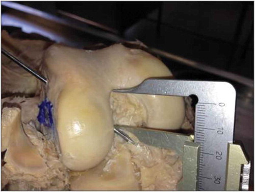
Figure 1. Guidewire passing between the anteromedial and posterolateral bundles of the ACL.
Source: Anatomy Laboratory–UNIRIO, 2015.
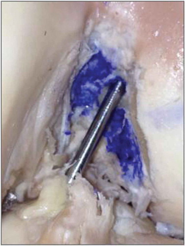
Figure 2. Ethibond 2 suture thread passing between the anteromedial and posterolateral bundles of the ACL.
Source: Anatomy Laboratory–UNIRIO, 2015.
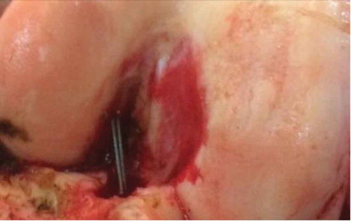
Figure 3. Hemostatic forceps placed on the suture thread, 1 cm from the tibial cortex with the knee in flexion.
Source: Anatomy Laboratory–UNIRIO, 2015.
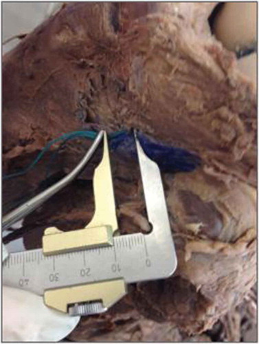
Figure 4. Thread measurement with the knee in extension.
Source: Anatomy Laboratory–UNIRIO, 2014–2015.
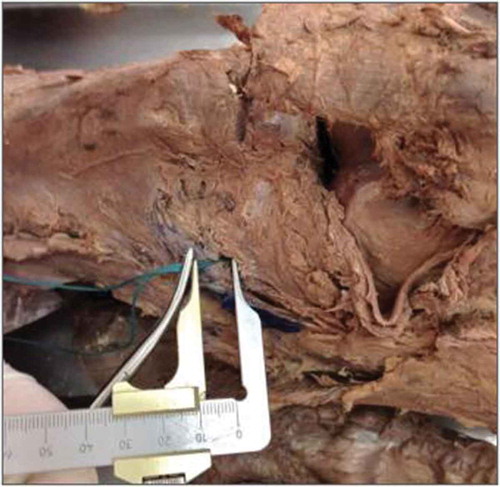
Figure 5. Distance from the guidewire placed in the tibial “footprint” to the PCL.
Source: Anatomy Laboratory–UNIRIO, 2014–2015.
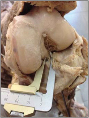
Figure 6. Height of the guidewire exit point in relation to the anteroposterior measurement of the lateral femoral condyle.
Source: Anatomy Laboratory–UNIRIO, 2014–2015.
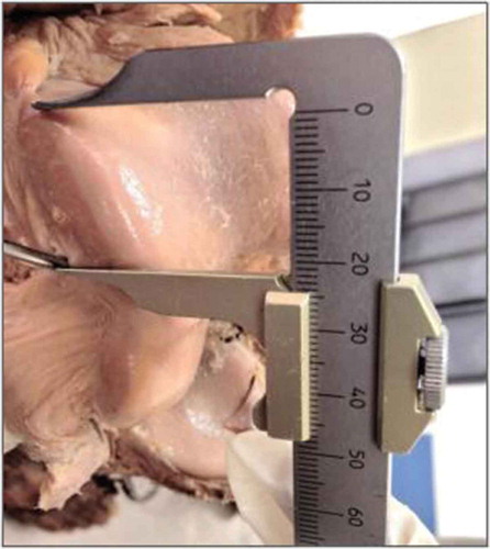
Figure 7. Height from the articular surface of the tibia to the intercondylar roof.
Source: Anatomy Laboratory–UNIRIO, 2014–2015.
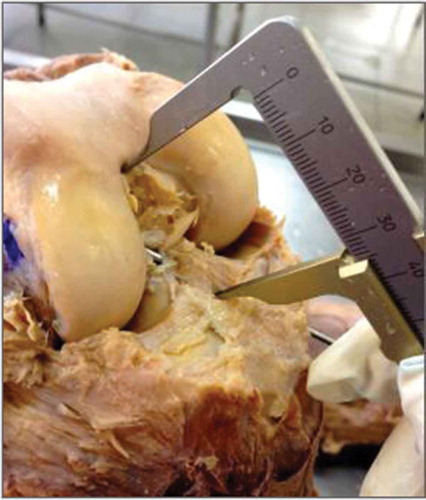
Table 1. Obtained values from eight measurements performed on the 16 studied knees
Table 2. Descriptive analysis of the distance between the guidewire and the intercondylar 341 roof and between the articular surface of the tibia and the femoral intercondylar roof 342