608
Views
1
CrossRef citations to date
0
Altmetric
Research Article

Faecal secretogranin and chromogranin levels persist over time and are unrelated to disease history and outcome in patients with ulcerative colitis

, , , , & | (Reviewing editor) show all
Article: 1484602 | Received 08 Dec 2017, Accepted 28 May 2018, Published online: 18 Jun 2018

Figures & data

Table 1. Demographics at baseline for patients with UC who stayed in remission (n = 20) or relapsed (n = 20) during the sample collection time

Figure 1. Faecal granin levels at baseline and relation to disease extent. Faecal samples from UC patients were obtained at baseline and analysed for secretogranins and chromogranins using RIA. (A) Levels of SN, Sg3, CgA and CgB for UC patients in remission (n = 40). (B) Comparison of levels of SN (left) and CgA (right) in UC patients with left-sided colitis (n = 25) and extensive colitis (n = 15). Each symbol represents one individual and horizontal lines indicate median of the group.
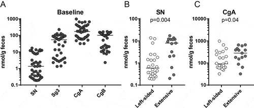
Figure 1. Faecal granin levels at baseline and relation to disease extent. Faecal samples from UC patients were obtained at baseline and analysed for secretogranins and chromogranins using RIA. (A) Levels of SN, Sg3, CgA and CgB for UC patients in remission (n = 40). (B) Comparison of levels of SN (left) and CgA (right) in UC patients with left-sided colitis (n = 25) and extensive colitis (n = 15). Each symbol represents one individual and horizontal lines indicate median of the group.

Figure 2. Faecal granin levels stay constant over time during remission. Faecal samples from UC patients were obtained at different time points and analysed for secretogranins and chromogranins using RIA. Levels of SN, Sg3, CgA and CgB for UC patients in remission were compared at three time points during a short interval (A) and at two time points during a long interval (B). Each symbol represents one individual (A: n = 10, B: n = 10).
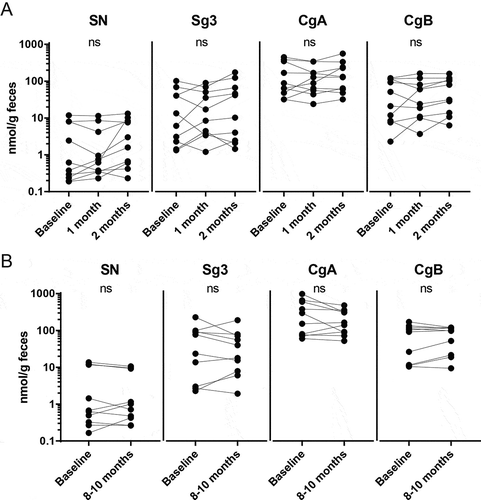
Figure 2. Faecal granin levels stay constant over time during remission. Faecal samples from UC patients were obtained at different time points and analysed for secretogranins and chromogranins using RIA. Levels of SN, Sg3, CgA and CgB for UC patients in remission were compared at three time points during a short interval (A) and at two time points during a long interval (B). Each symbol represents one individual (A: n = 10, B: n = 10).

Figure 3. Faecal granin levels stay constant over time before, during and after a relapse. Faecal samples from UC patients were obtained at different time points and analysed for calprotectin, secretogranins and chromogranins using ELISA and RIA. Levels of calprotectin (A) and SN, Sg3, CgA and CgB (B) for UC patients 1–3 months before, during and 2–12 months after a relapse. Each symbol represents one individual (n = 20). ****p-value < 0.0001 and ***p-value < 0.001.
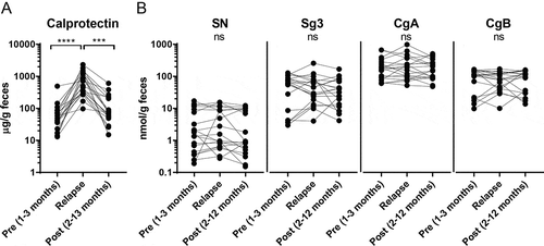
Figure 3. Faecal granin levels stay constant over time before, during and after a relapse. Faecal samples from UC patients were obtained at different time points and analysed for calprotectin, secretogranins and chromogranins using ELISA and RIA. Levels of calprotectin (A) and SN, Sg3, CgA and CgB (B) for UC patients 1–3 months before, during and 2–12 months after a relapse. Each symbol represents one individual (n = 20). ****p-value < 0.0001 and ***p-value < 0.001.

Table 2. Correlations between SN, Sg3, CgA and CgB levels in faecal samples obtained at baseline1

Figure 4. UC patients cluster into three different groups dependent on faecal granin levels. Median levels of faecal SN, Sg3, CgA and CgB from three to seven samples were used for principle component analysis (PCA) and hierarchical clustering analysis (HCA). Score scatter plot (A), HCA (B) and loading scatter plot (C) for the PCA are shown. The shapes from the groups defined by the HCA are highlighted in (A). Each symbol in A represents one individual. Patients are numbered 10–49.
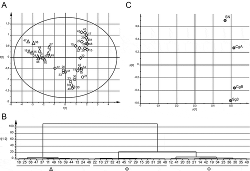
Figure 4. UC patients cluster into three different groups dependent on faecal granin levels. Median levels of faecal SN, Sg3, CgA and CgB from three to seven samples were used for principle component analysis (PCA) and hierarchical clustering analysis (HCA). Score scatter plot (A), HCA (B) and loading scatter plot (C) for the PCA are shown. The shapes from the groups defined by the HCA are highlighted in (A). Each symbol in A represents one individual. Patients are numbered 10–49.

Table 3. Relation between faecal granin expression and disease outcome

Supplemental material

Supplemental Material

Download ()