Figures & data
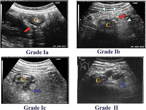
Figure 1. Classification of ACD on the basis of US. The severity of ACD was classified into two grades. Grade Ia: an inflamed diverticulum (arrow). “C” indicates colon. Grade Ib: an inflamed diverticulum (arrow) with pericolitis (arrow head). Grade Ic: an inflamed diverticulum with abscess formation of ≤ 2 cm in diameter (arrow). Grade II: an inflamed diverticulum with abscess formation of > 2 cm in diameter (arrow) or with perforation.
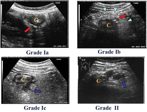
Table 1. Patient characteristics
Table 2. Results of grades of acute colon deverticulitis between ultrasound sonography (US) and computer tomography (CT)
Figure 2. Comparison of findings by ultrasonography (US) with those by computed tomography (CT).
(A) In more than 60% of cases, US Grades Ia were diagnosed as Grade Ia. The red arrow indicates the inflamed diverticulum. The arrow indicates the inflamed diverticulum. The arrowhead indicate the inflamed pericolic tissue. The letter “C” indicates colon.(B) In more than 90% of cases, US Grades Ib were diagnosed as Grade Ib. The arrow indicates the inflamed diverticulum. The arrowheads indicate the inflamed pericolic tissue. The letter “C” indicates colon.(C) In more than 25% of cases, US Grades Ic were diagnosed as Grade Ic. The arrow indicates the small abscess. The arrowhead indicates the inflamed diverticulum. The letter “C”indicates colon.Da,b) In more than 50% of cases, US Grades II were diagnosed as Grade II. The long arrow indicates a large abscess. The short arrow indicated free air. The arrowhead indicates air leakage. The letter “C” indicates colon.
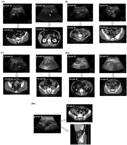
Table 3. Patients characteristics of outpatient treatment
Table 4. Clinical results in Right and Left side ACD patients