
Figures & data
Figure 1. (A) Preoperative X-ray. (B) Postoperative X-ray (immediately after ulnar shortening osteotomy). (C) Postoperative X-ray (3 months after the surgery). (D) Postoperative X-ray (5 months after the surgery). The distal screw was loosened (Arrow). There was a bony absorption at the osteotomy site.
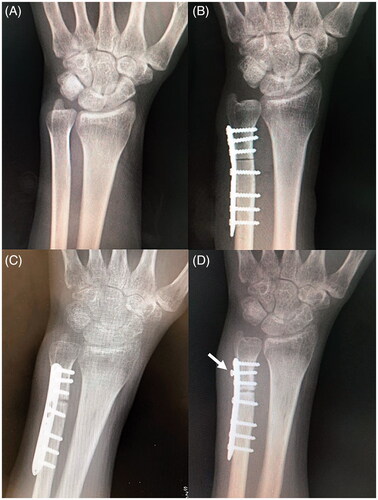
Figure 2. (A) Postoperative X-ray (immediately after implant removal and debridement). (B) Postoperative computed tomography (CT, 1 month after implant removal and debridement). There was poor callus formation and a bony absorption at the osteotomy site. (C) Postoperative CT (4 months after implant removal and debridement and 3 months after the start of teriparatide therapy and low-intensity pulsed ultrasound [LIPUS]). (D) Postoperative CT (5 months after implant removal and debridement and 4 months after the start of teriparatide therapy and LIPUS). Bony union at the osteotomy site was observed.
![Figure 2. (A) Postoperative X-ray (immediately after implant removal and debridement). (B) Postoperative computed tomography (CT, 1 month after implant removal and debridement). There was poor callus formation and a bony absorption at the osteotomy site. (C) Postoperative CT (4 months after implant removal and debridement and 3 months after the start of teriparatide therapy and low-intensity pulsed ultrasound [LIPUS]). (D) Postoperative CT (5 months after implant removal and debridement and 4 months after the start of teriparatide therapy and LIPUS). Bony union at the osteotomy site was observed.](/cms/asset/7087d3c0-f8ed-409d-83e7-7b797b49ee15/icrp_a_1894155_f0002_c.jpg)