Figures & data
Figure 1. Expression of PGT mRNA in the subarachnoidal space (a, b, c) and choroid plexus (d, e) of the brain and in the subarachnoidal space of the spinal cord (f, g). (A, D, F) 5 h after saline injection, (B, E, G) 5 h after LPS injection, (C) hybridization with sense probe (5 h after LPS injection). PGT mRNA was expressed in the arachnoid membrane (arrowheads), subarachnoidal blood vessels (arrows), and blood vessels in the choroid plexus (asterisk) after the LPS injection. A line drawing at the upper right corner indicates brain regions corresponding to a–e. Scale bar: 50 µm.
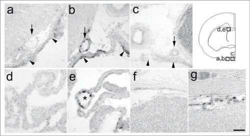
Figure 2. Time-dependent expression of PGT mRNA in the brain subarachnoidal space. (A) untreated rat, (B) 1.5 h, (C) 3 h, (D) 5 h, (E) 12 h, (F) 24 h, (G) 48 h after the LPS injection. (H) 5 h after the saline injection. Levels of PGT mRNA in the arachnoid membrane (arrowheads) and subarachnoidal blood vessels (arrows) reached their peak at 5 h after the LPS injection. Inlets show 3-times magnified views of blood vessels. Scale bar: 100 µm.
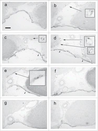
Figure 3. Semiquantitative analysis of PGT mRNA. (A) changes in the number of PGT mRNA-positive cells in blood vessels in the subarachoidal space after LPS injection, (B) changes in signal intensity of PGT mRNA in the arachnoid membrane after LPS injection. The values denoted by asterisks were significantly different from that of UT (untreated) group (*P < 0.05, **P < 0.01, ***P < 0.001).
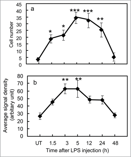
Figure 4. Expression of PGT protein. (A) Western blot of the rat brain (5 h after the LPS injection) with (+) or without (−) PGT antibody. (B–D) confocal microscopic views of the lung of saline-injected rat. (B) expression of PGT protein in the lung. (C) a magnified view of “b.” PGT is expressed in a vesicular pattern. (D) PGT antibody was incubated with the antigen peptide prior to assay. (E–H) confocal microscopic views of the brain sampled at 12 h after the LPS injection. (E) expression of PGT protein in the arachnoid membrane. (F) arachnoid membrane when incubated with the preabsorbed PGT antibody. (G) a magnified view of a blood vessel, which expressed PGT in vesicular pattern. The asterisk indicates the lumen of a blood vessel. (H) a magnified view of a blood vessel when incubated with the preabsorbed PGT antibody. Scale bars: 100 µm (b,d), 50 µm (e,f), 5 µm (c,g,h).
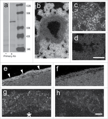
Figure 5. Expression of PGT protein in brain subarachnoidal space at each time point after LPS or saline injection. (A) 5 h after the saline injection (B) 5 h, (C) 12 h, (D) 24 h, (E) 48 h after the LPS injection. PGT was induced in venous blood vessels (asterisks) after the LPS injection. Scale bar: 20 mm (F) semiquantitative analysis of the signal intensity of PGT protein at 24 h after the LPS injection (closed bar) and 5 h after the saline injection (open bar). *P < 0.05.
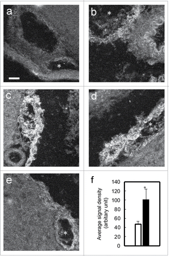
Figure 6. PGT protein localization in blood vessels. (A–C) confocal microscopic views of a venous blood vessel. (A) expression of PGT protein (B) expression of von Willbrand Factor, an endothelial marker. (C) double-stained image of PGT and von Willbrand Factor. PGT was expressed in endothelial cells of the venous blood vessels (whose lumen is indicated by the asterisk) and arachnoid membrane (arrowhead). (D, F) COX-2 protein expression in a subarachnoidal blood vessel (d) and a parenchymal blood vessel (f). (E, G) PGT mRNA expression in subarachnoidal blood vessel (e) in an adjacent section to (D) and in a parenchymal blood vessel (g) in an adjacent section to (F). PGT mRNA and COX-2 were coexpressed in subarachnoidal venous blood vessels. In parenchymal blood vessels, little PGT mRNA was expressed, whereas, COX-2 was expressed well. Scale bar: 20 µm.
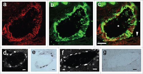
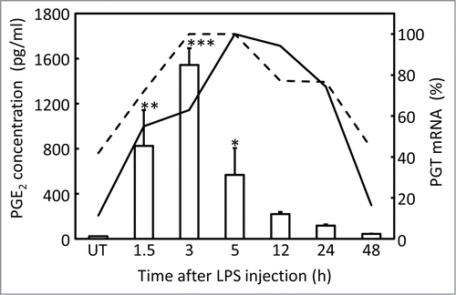
Figure 7. Time-course analysis of PGE2 concentration in the CSF (open columns), and PGT mRNA in the brain (subarachnoidal blood vessels: solid line, arachnoid membrane: dotted line) after LPS injection. Values of PGT mRNA signals were converted to a percentage of the maximum level obtained at 5 h after the injection. The original values and SE were shown in . PGE2 concentration denoted by asterisks were significantly different from that of UT (untreated) group (*P < 0.05, **P < 0.01, ***P < 0.001).